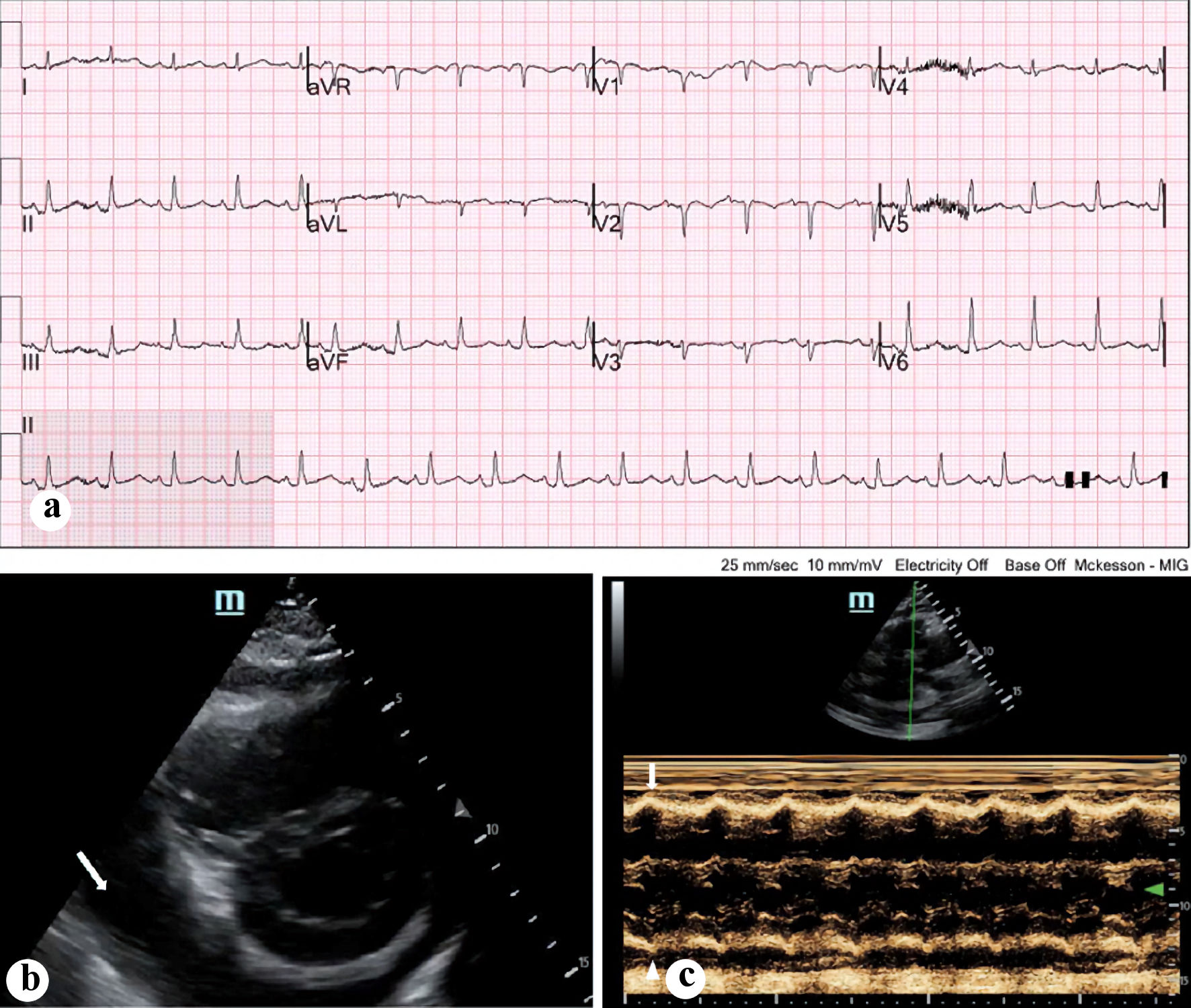
Figure 1. (a) Electrocardiogram done on presentation, demonstrating sinus tachycardia with relatively low voltages. (b) Bedside echocardiogram done on presentation, with a parasternal view demonstrating circumferential pericardial effusion (arrow). (c) M-mode ultrasonography demonstrating early right ventricular collapse during diastole (arrow).
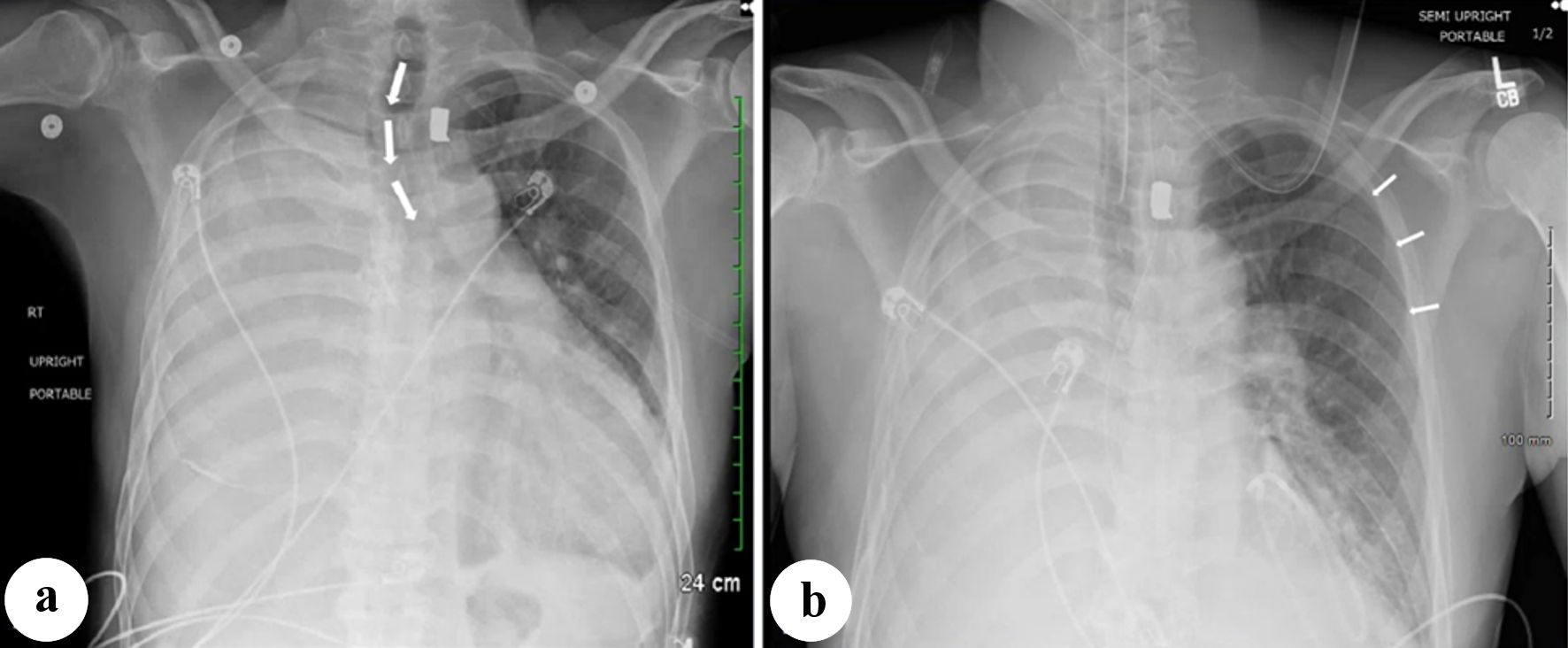
Figure 2. (a) Portable chest X-ray done on presentation, demonstrating right-sided white-out and cardiomegaly along with shift in the mediastinal airway structure (arrows). (b) Significant improvement in left lung volume following pericardiocentesis (arrows).
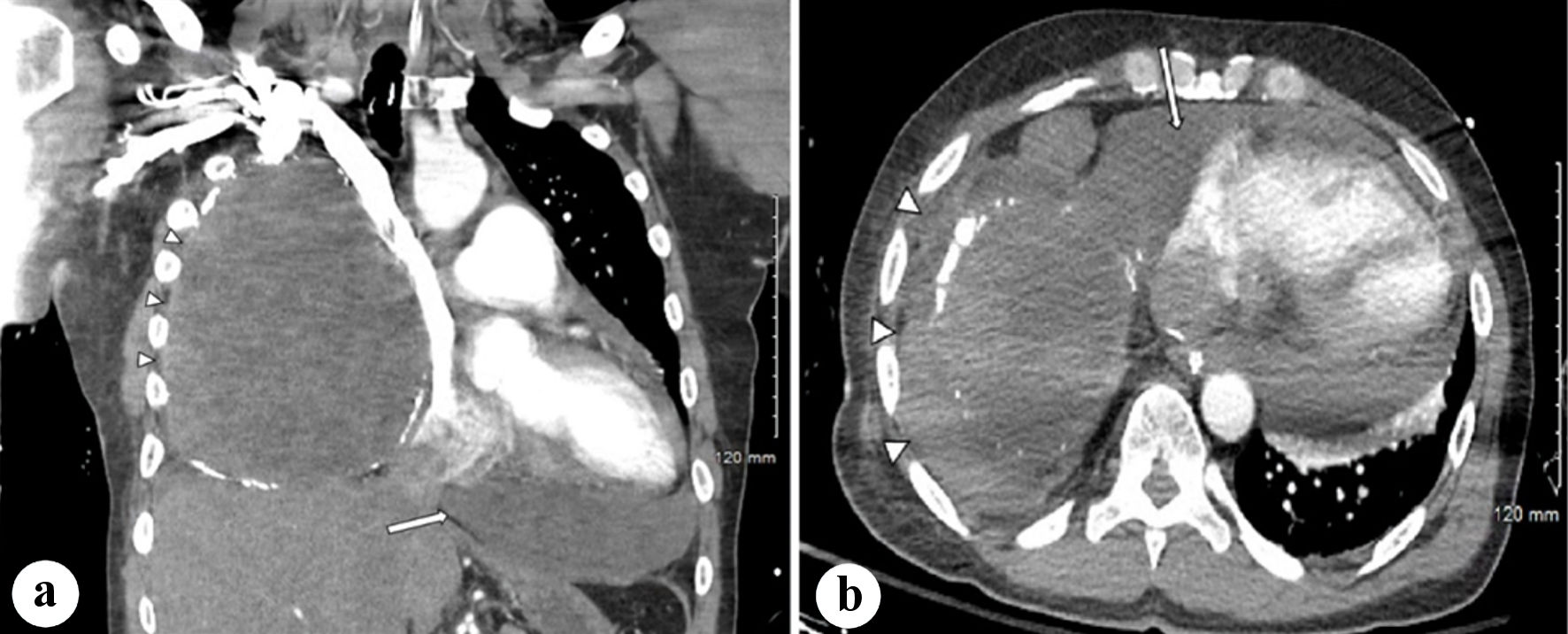
Figure 3. (a) Coronal CT scan of the thorax with IV contrast demonstrating moderate-large pericardial effusion and fluid collection within the right thorax with surrounding calcifications, with mediastinal shift (arrows). (b) Axial CT scan of the thorax with IV contrast demonstrating right-sided fluid collection with calcifications as well as the pericardial effusion (arrows). CT: computed tomography; IV: intravenous.