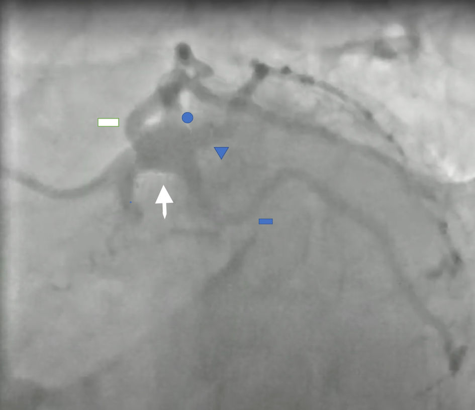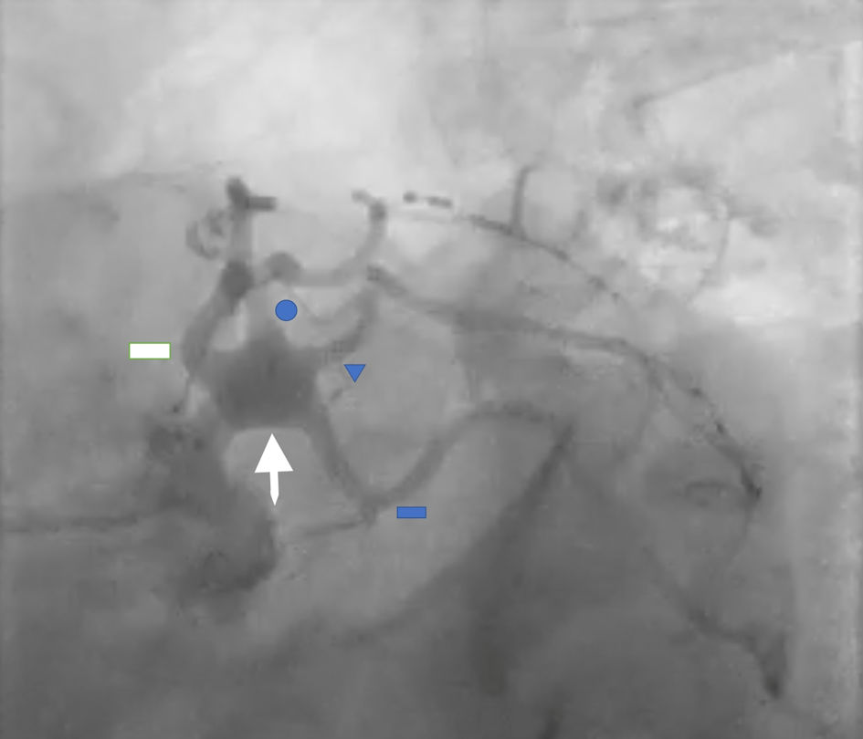
Figure 1. Left anterior oblique caudal (LAO caudal) view showed aneurysmal left main (LM) coronary artery (white arrow) with four branches (quadfurcation). Blue circle: first ramus intermedius artery, blue triangle: second ramus intermedius artery, angiographically mild luminal irregularities of the left anterior descending, first and second ramus intermedius, and left circumflex coronary arteries. White rectangle: left anterior descending artery; blue rectangle: left circumflex artery.