Figures
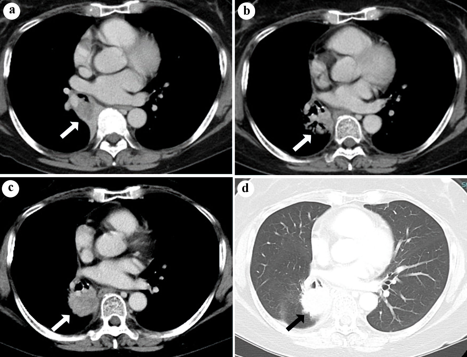
Figure 1. Chest CT (a) in October 2019, before chemo-radiation, (b) in January 2022, 1 year after durvalumab and (c, d) in July 2022, 1.5 years after durvalumab. The white (a-c) or black (d) arrows indicate the shadow of the primary tumor (a, b) or sarcoid-like granulomatosis (c, d) in the right lower lobe. CT: computed tomography.
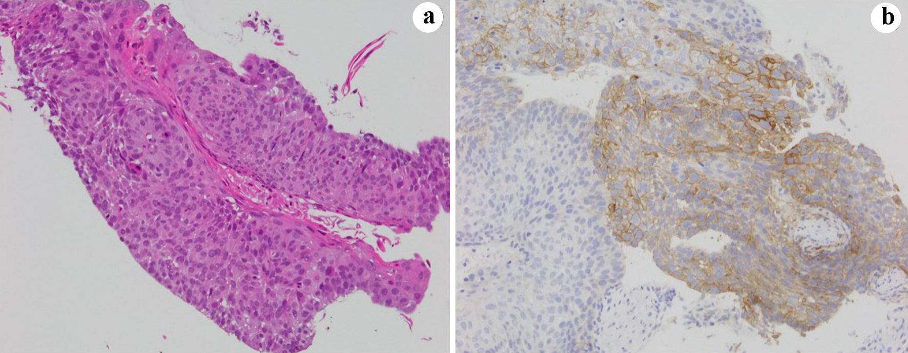
Figure 2. Bronchoscopic sample in October 2019 at the diagnosis. (a) Hematoxylin and eosin and (b) PD-L1 (22C3 pharmDx assay) stains. PD-L1: PD-1 ligand; PD-1: programmed cell death protein 1.
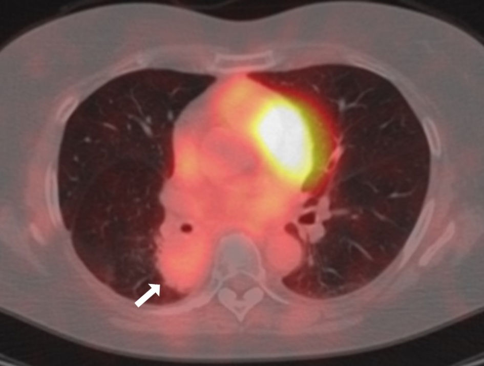
Figure 3. Fused image of fluorodeoxyglucose positron emission tomography/computed tomography (FDG-PET/CT) in July 2022 before the right lower lobe lobectomy. The white arrow indicates the sarcoid-like granulomatosis lesion. FDG-PET: fluorodeoxyglucose positron emission tomography; CT: computed tomography.
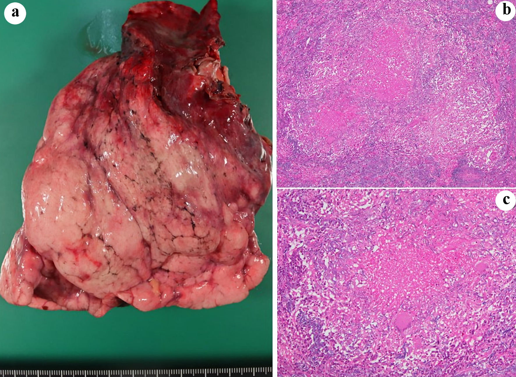
Figure 4. Macroscopical (a) and histopathological (b, c) findings of the resected right lower lobe. (b, c) Victoria blue/hematoxylin and eosin stain (magnification; (b) × 4, (c) × 10).
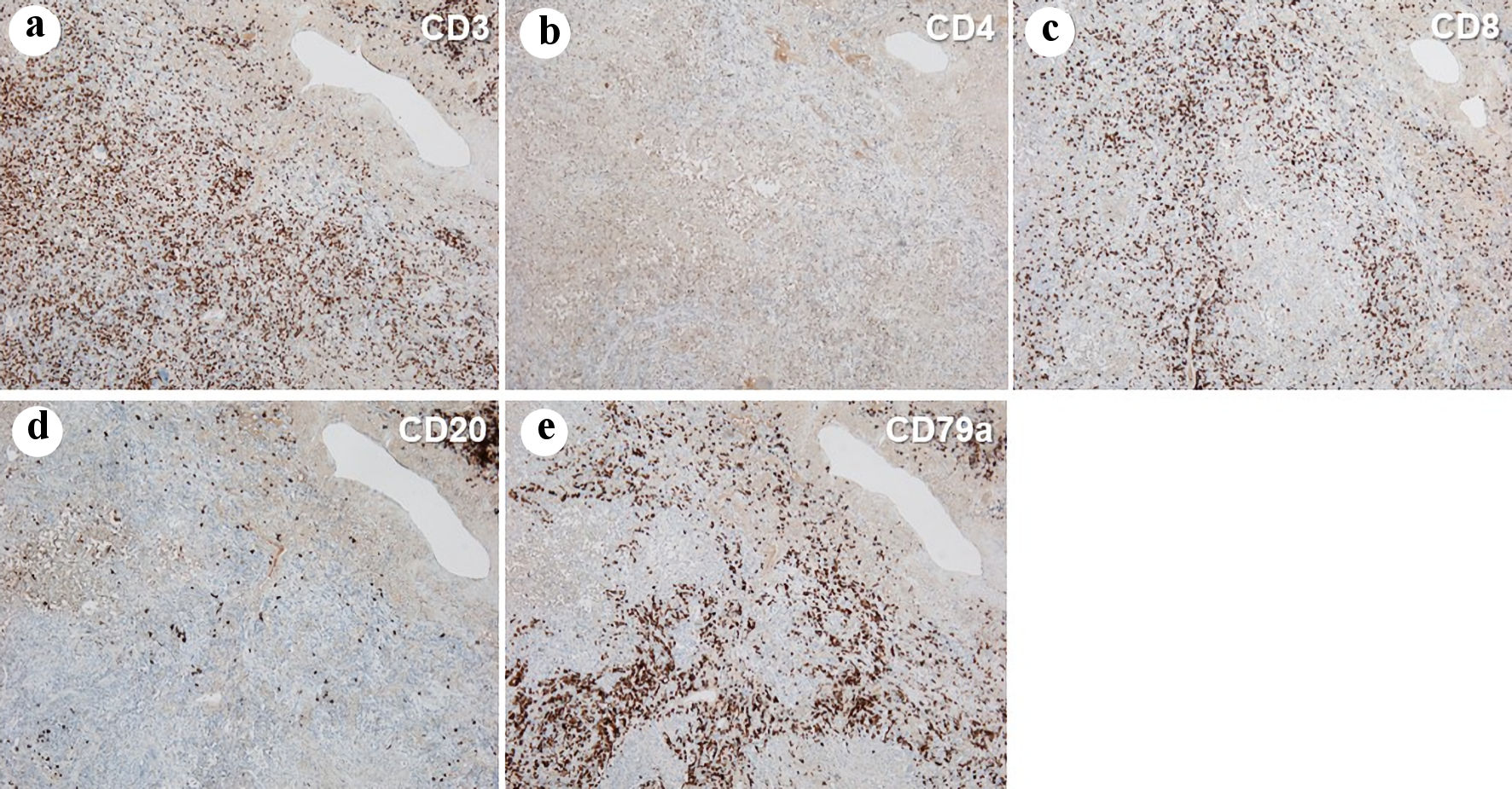
Figure 5. Immunohistochemistry of the resected right lower lobe, stained by anti-CD3 (a), CD4 (b), CD8 (c), CD20 (d) and CD79a (e) antibodies (magnification: × 4).