Journal of Medical Cases, ISSN 1923-4155 print, 1923-4163 online, Open Access
Article copyright, the authors; Journal compilation copyright, J Med Cases and Elmer Press Inc
Journal website https://www.journalmc.org

Case Report

Volume 14, Number 5, May 2023, pages 155-161


Boon or Bane? Anti-Tumor Necrosis Factor Therapy Complicated by Listeria monocytogenes Meningitis Culminating in Colectomy for Ulcerative Colitis

Figures

Figure 1.
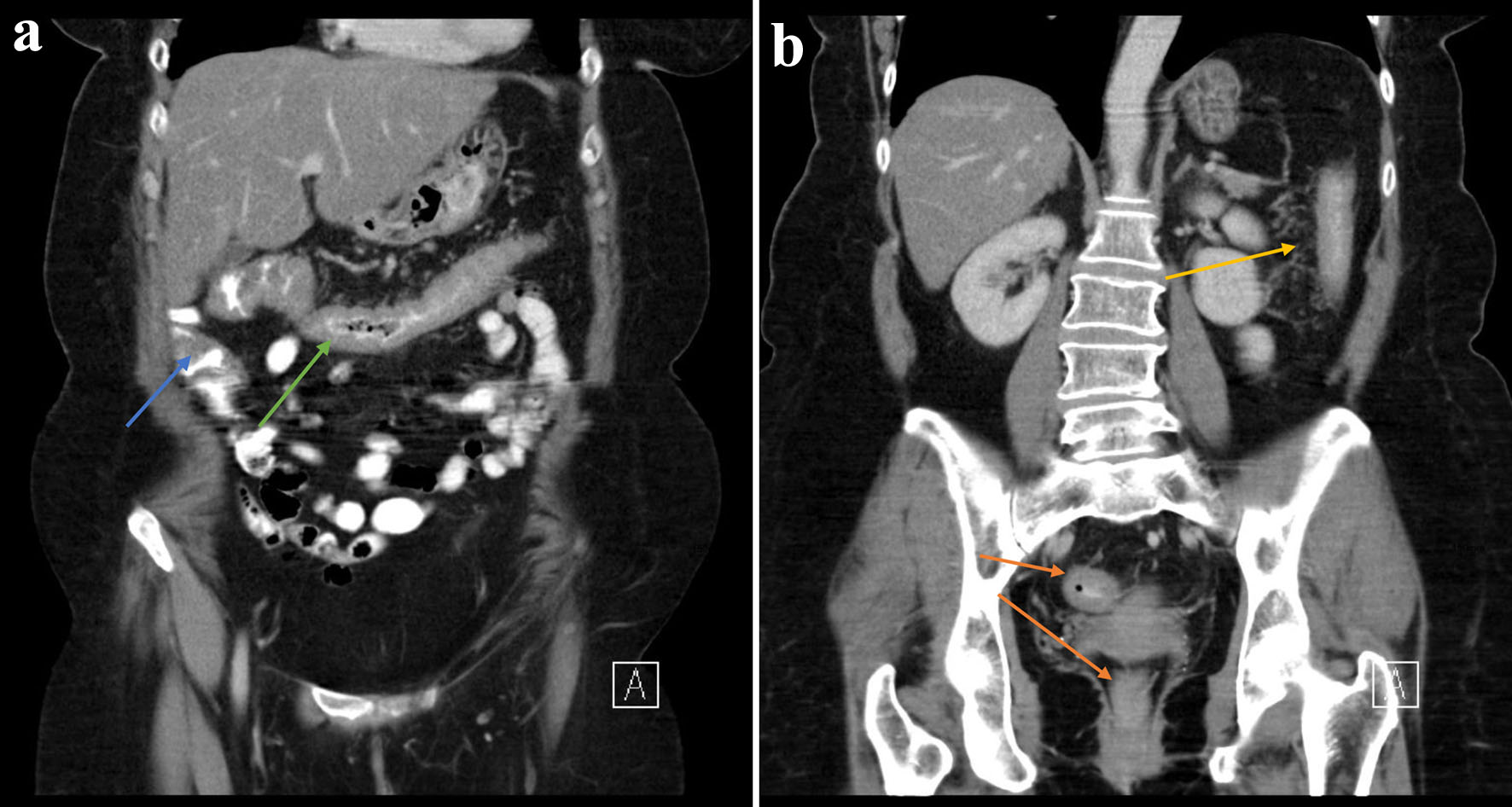
Figure 1. (a, b) There is circumferential thickening of the distal ascending (blue arrow), transverse (green arrow), descending (yellow arrow) and rectosigmoid colon (orange arrow) in keeping with colitis.
Figure 2.
Figure 2. Computed tomography of brain (09/10/2021) demonstrating normal parenchymal appearances. Lumbar puncture revealed grossly cloudy cerebrospinal fluid with massively elevated white blood cell count of 1,600/mm3, with 70% polymorphonuclear neutrophils and 30% mononuclear cells. No organisms were seen on gram stains with no growth on cultures but Listeria monocytogenes DNA was detected via polymerase chain reaction. Cerebrospinal fluid (CSF) glucose was low at 1.8 mmol/L (2.22 to 4.44) and CSF protein was raised at 1.75 g/L (0.15 to 0.6) which confirmed a bacterial meningitis. Apart from bacteremia and meningitis, there were no other manifestations of listeriosis in this patient.
Figure 3.
Figure 3. Endoscopic photograph of sigmoid - edematous mucosa (blue arrow), erosions (green arrow) and ulcerations (grey arrow). Also note absence of vascular pattern with marked erythematous appearances.