Journal of Medical Cases, ISSN 1923-4155 print, 1923-4163 online, Open Access
Article copyright, the authors; Journal compilation copyright, J Med Cases and Elmer Press Inc
Journal website https://www.journalmc.org

Case Report

Volume 14, Number 3, March 2023, pages 95-99


Fracture Neck of the Femur: A Case of Two Pathologies

Figures

Figure 1.
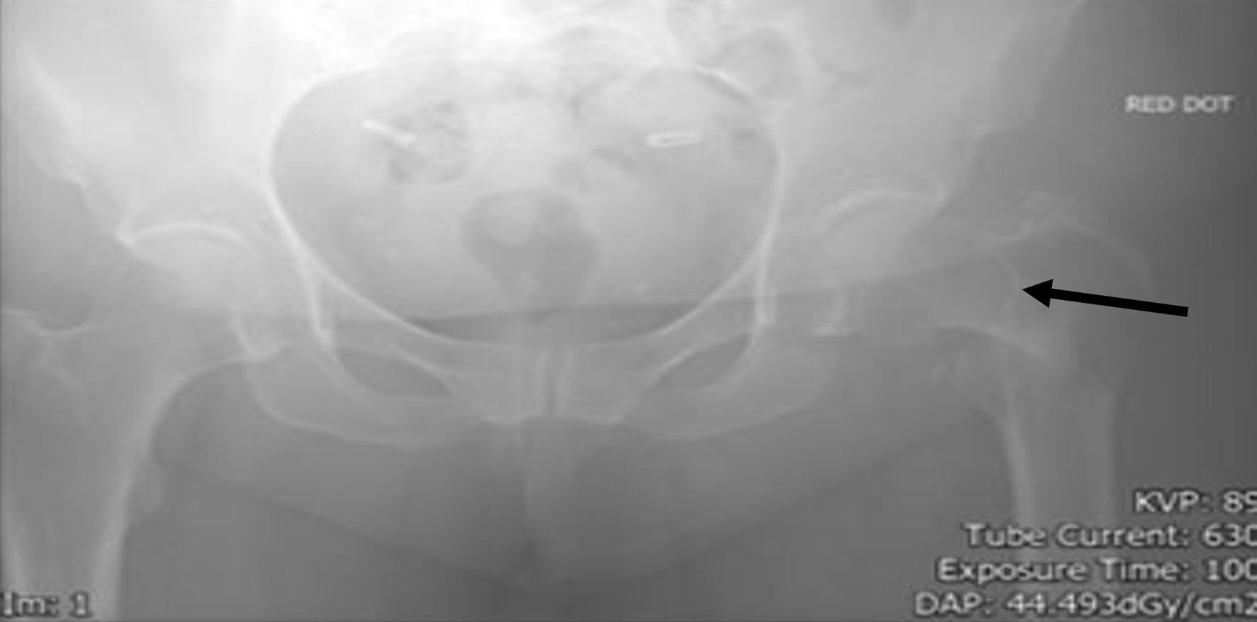
Figure 1. The pelvic X-ray on admission (no clear evidence of fracture of the left neck of the femur).
Figure 2.
Figure 2. The repeat of pelvic X-ray, 4 days after the X-ray in Figure 1. Arrow points to the site of the fracture of the left neck of the femur.
Figure 3.
Figure 3. Computed tomography (CT) scan showing the enlargement of the thyroid gland (blue arrow points to the enlarged thyroid). The rest of the CT scan of the neck, abomen and plevis showed no other abnormalities.
Figure 4.
Figure 4. PET scan and high uptake can be seen in the right lobe of thyroid gland. The conclusion of all the investigations: this diffuse B-cell lymphoma is confined to thyroid gland and left neck of the femur.