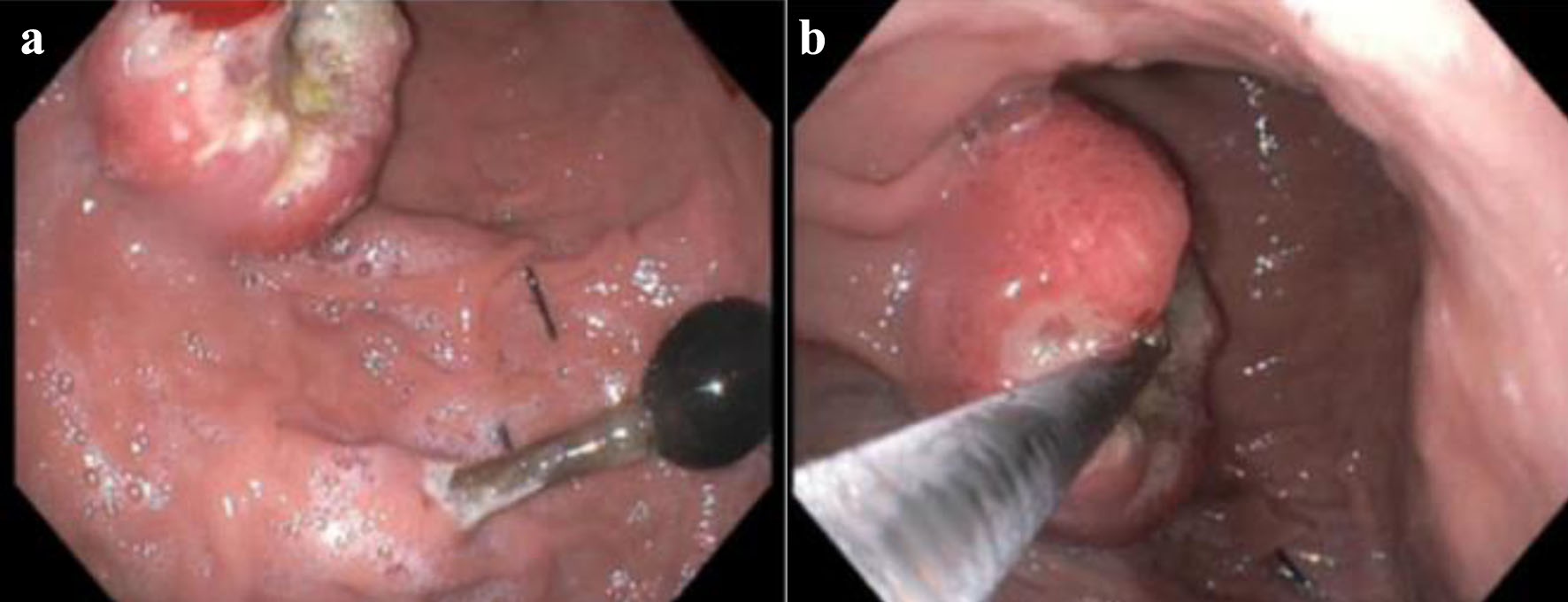
Figure 1. (a) The proximal end of PEG. (b) EGD showing an ulcerating circumferential gastric mass at the gastric body. PEG: percutaneous endoscopic gastrostomy; EGD: esophagogastroduodenoscopy.
| Journal of Medical Cases, ISSN 1923-4155 print, 1923-4163 online, Open Access |
| Article copyright, the authors; Journal compilation copyright, J Med Cases and Elmer Press Inc |
| Journal website https://www.journalmc.org |
Case Report
Volume 14, Number 3, March 2023, pages 100-104
Metastatic Squamous Cell Carcinoma From Primary Hypopharynx Source to Gastric Mucosa Presenting as Massive Gastrointestinal Bleeding
Figures