Journal of Medical Cases, ISSN 1923-4155 print, 1923-4163 online, Open Access
Article copyright, the authors; Journal compilation copyright, J Med Cases and Elmer Press Inc
Journal website https://www.journalmc.org

Case Report

Volume 14, Number 3, March 2023, pages 105-110


Subarachnoid Hemorrhage From Cavernous Malformation Masquerading as Myocardial Infarction

Figures

Figure 1.
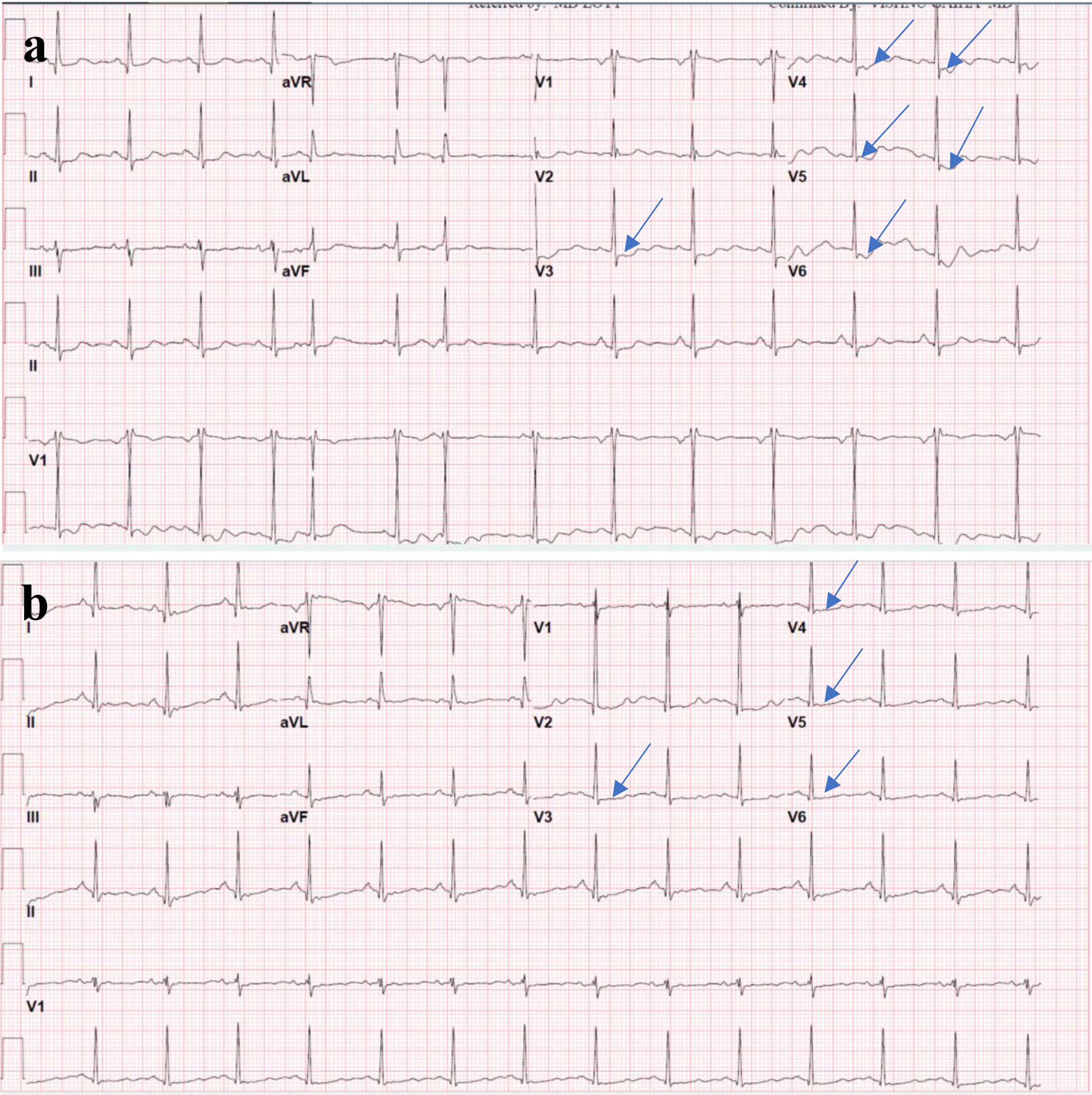
Figure 1. (a) ECG on presentation showing ST segment depression (arrows) in the anterolateral leads (V3-V6). (b) Repeat ECG on the next day showing improvement in the ST segment deviations noticed on the initial ECG. ECG: electrocardiogram.
Figure 2.
Figure 2. (a, b) Negative coronary angiography. (c) A single axial CT angiogram of the chest without a pulmonary artery filling defect, negative for pulmonary embolus. (d) A sagittal slice of a CT aortogram negative for aortic dissection. CT: computed tomography.
Figure 3.
Figure 3. (a) An axial cut of a brain CT demonstrating acute SAH (arrow) in the left occipital lobe. (b) An axial cut of a CT brain demonstrating resolving SAH (arrow) on discharge. (c) The outpatient follow-up head scan at 3 weeks showing a resolved SAH and a focal hyperdensity (arrow) which raised suspicion for arteriovenous malformation. (d) A follow-up MRI 5 weeks after discharge showing a sub-centimeter cavernoma (arrow) adjacent to resolving SAH blood products. CT: computed tomography; SAH: subarachnoid hemorrhage; MRI: magnetic resonance imaging.