Journal of Medical Cases, ISSN 1923-4155 print, 1923-4163 online, Open Access
Article copyright, the authors; Journal compilation copyright, J Med Cases and Elmer Press Inc
Journal website https://www.journalmc.org

Case Report

Volume 14, Number 5, May 2023, pages 162-168


Vanishing Bile Duct Syndrome in a Patient With Recurrent Hodgkin Lymphoma

Figures

Figure 1.
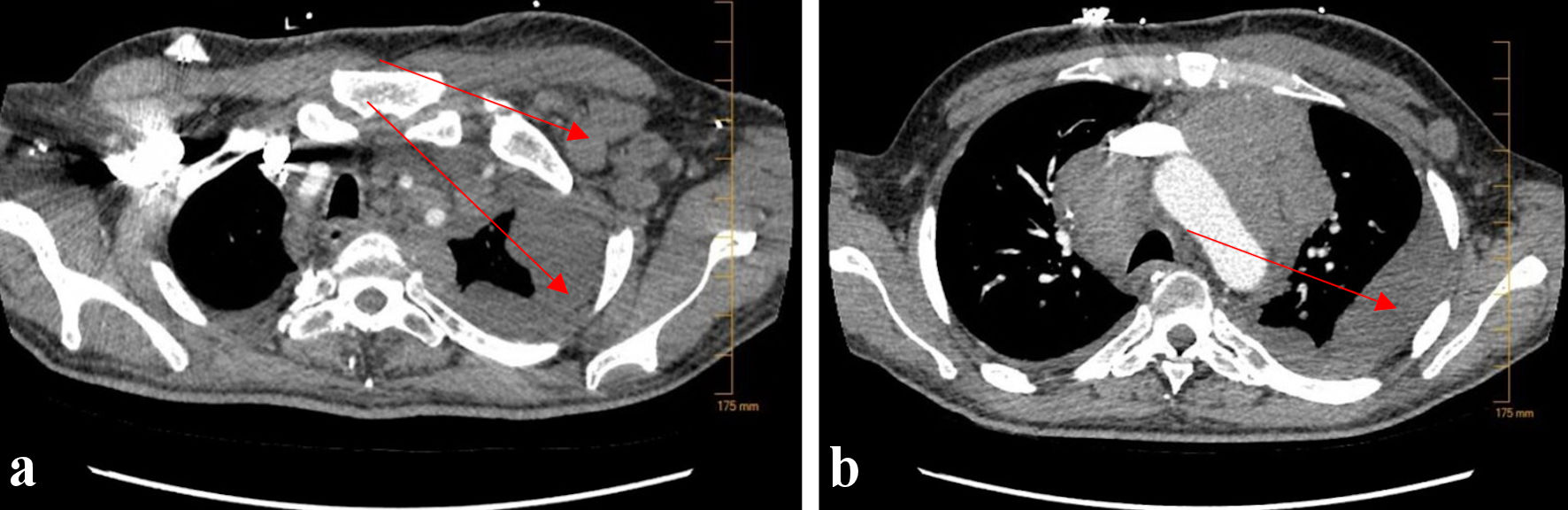
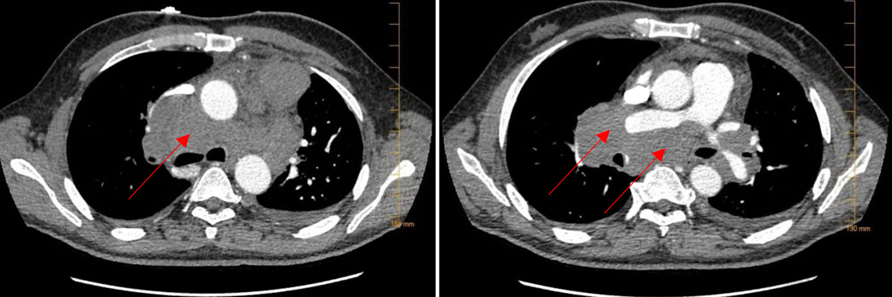
Figure 1. CTA of the lungs. CTA of the lungs on admission showed extensive bulky mediastinal and hilar lymphadenopathy with encasement of the pulmonary artery (arrows). CTA: computed tomography angiography.
Figure 2.
Figure 2. CT abdomen/pelvis with contrast. (a) CT abdomen/pelvis demonstrated a normal appearing liver without evidence of biliary tract dilation. (b) Additional bulky lymphadenopathy was also noted in the aortocaval node region as well as para-diaphragmatic nodes (arrow). CT: computed tomography.
Figure 3.
Figure 3. CTA of the lungs upon re-admission 2 months later. Repeat CTA of the lungs showed new, bulky left-sided axillary lymphadenopathy (a) (arrow), as well as extensive, worsening left-sided pleural effusion (a, b) (arrows). CTA: computed tomography angiography.
Figure 4.
Figure 4. Still image from ERCP study demonstrating failure of contrast penetration through the biliary tree. Findings additionally demonstrate atrophy of intrahepatic ducts with appearance of sclerosing cholangitis (arrow). ERCP: endoscopic retrograde cholangiopancreatography.
Figure 5.
Figure 5. Liver biopsy demonstrating absence of interlobular bile duct and hepatocyte injury, consistent with VBDS (arrow). VBDS: vanishing bile duct syndrome.