Journal of Medical Cases, ISSN 1923-4155 print, 1923-4163 online, Open Access
Article copyright, the authors; Journal compilation copyright, J Med Cases and Elmer Press Inc
Journal website https://www.journalmc.org

Case Report

Volume 14, Number 4, April 2023, pages 141-147


A Case Series of Non-Tuberculous Mycobacterial Pulmonary Disease Masquerading as Malignancy From a Community-Based Hospital

Figures

Figure 1.
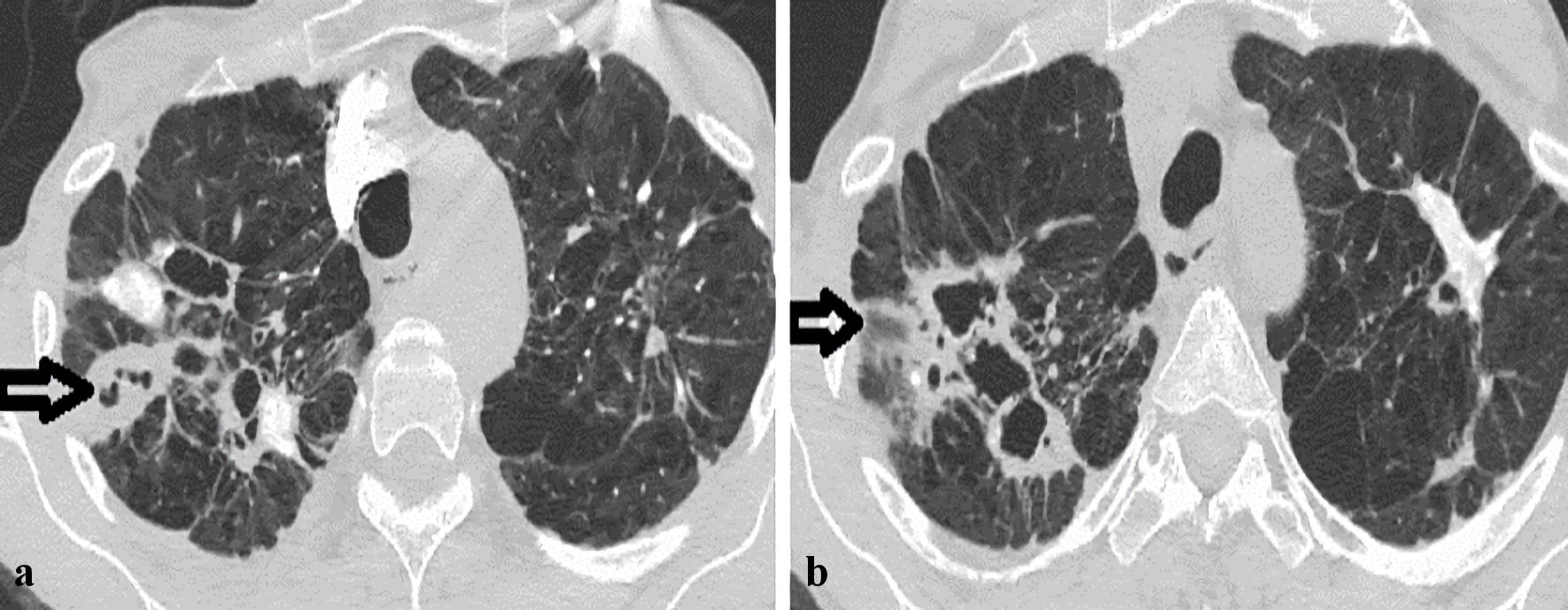
Figure 1. Axial images of computed tomography (CT) scans of the chest demonstrating interval progression of the left-sided lung nodules in the upper lobe over a period. (a) Previous CT chest scan 2 years prior to presentation. (b) CT chest scan on index presentation (arrow and pointer showing interval nodule size progression).
Figure 2.
Figure 2. Axial images of computed tomography (CT) scans of the chest demonstrating interval progression of the right upper cavitary over 1 year (horizontal arrows). (a) CT chest scan on presentation. (b) Previous CT chest scan over 1 year prior to presentation.
Figure 3.
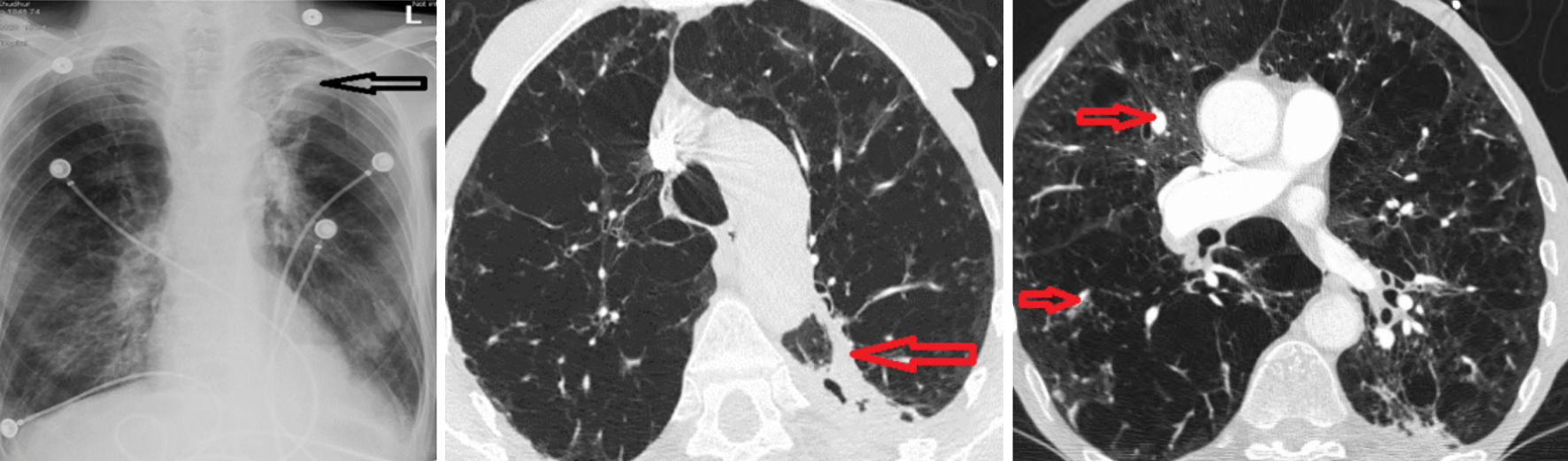
Figure 3. (a) Chest X-ray with the reticulonodular process and a loss of volume in the left upper lobe (horizontal arrow). (b) Contrast-enhanced CT chest revealed a cavitary lesion in the posterior aspect of the left upper lobe (LUL) with a solid component (horizontal arrow). (c) Diffuse nodular lesions (small horizontal arrows).