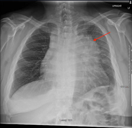
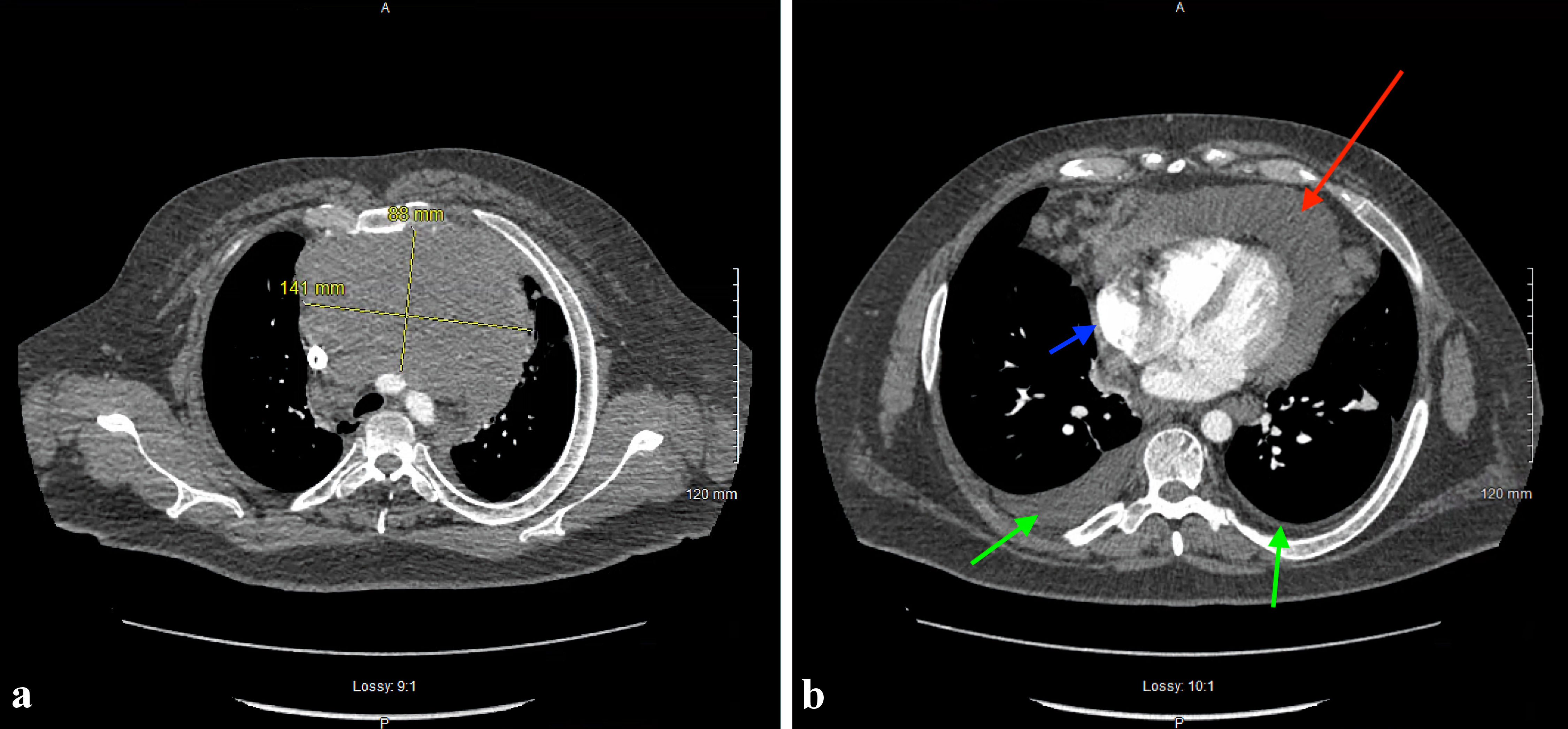
Figure 1. Posterior-anterior chest radiograph demonstrating a density bordering the aortic arch, descending thoracic aorta, and upper portion of the left heart (red arrow).
| Journal of Medical Cases, ISSN 1923-4155 print, 1923-4163 online, Open Access |
| Article copyright, the authors; Journal compilation copyright, J Med Cases and Elmer Press Inc |
| Journal website https://www.journalmc.org |
Case Report
Volume 14, Number 8, August 2023, pages 277-281
Primary Mediastinal B-Cell Lymphoma Presenting as Cardiac Tamponade
Figures
Table
| Laboratory study | Reference values | Measured values |
|---|---|---|
| White blood cells (cells/µL) | 4,300 - 11,100 | 9,400 |
| Hemoglobin (g/dL) | 13,000 - 17,000 | 12,600 |
| Mean corpuscular volume (fL) | 80 - 100 | 85 |
| Platelet (cells/µL) | 120,000 - 360,000 | 398,000 |
| Neutrophils (%) | 38 - 75 | 83 |
| Lymphocytes (%) | 20 - 48 | 6% |
| D-dimer (ng/mL) | 0 - 250 | 1,595 |
| Troponin-I (ng/mL) | 0 - 0.3 | < 0.3 |