Figures
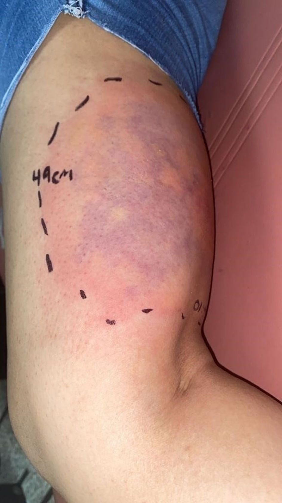
Figure 1. Lesion located on the right pelvic limb, with edema, erythema, blue-gray halo, irregular margins, with an approximate diameter of 49 cm.
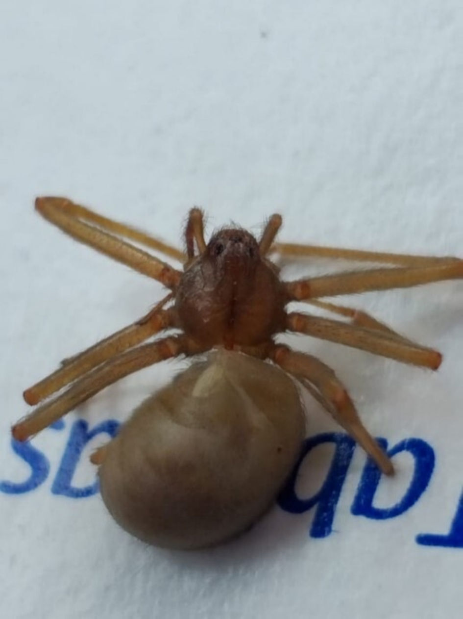
Figure 2. Arachnid of the genus Loxosceles presented by the patient.
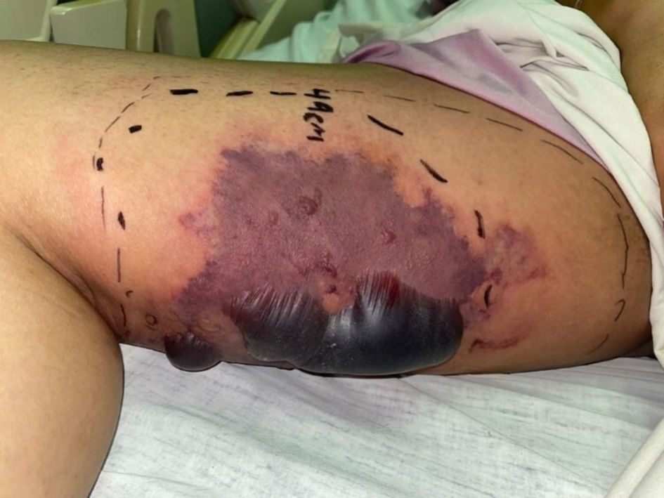
Figure 3. Decrease of erythema in the lesion, consolidation of liveloid plaque, and formation of vesicles with serohematous content.
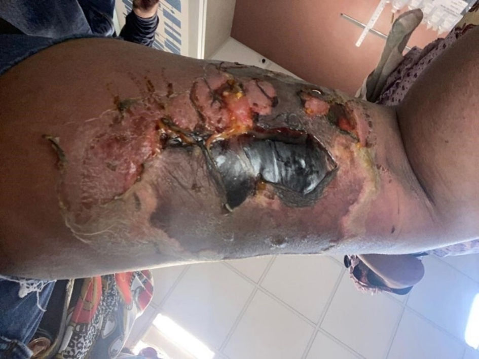
Figure 4. Rupture of the vesicles and desquamation of the dermis.
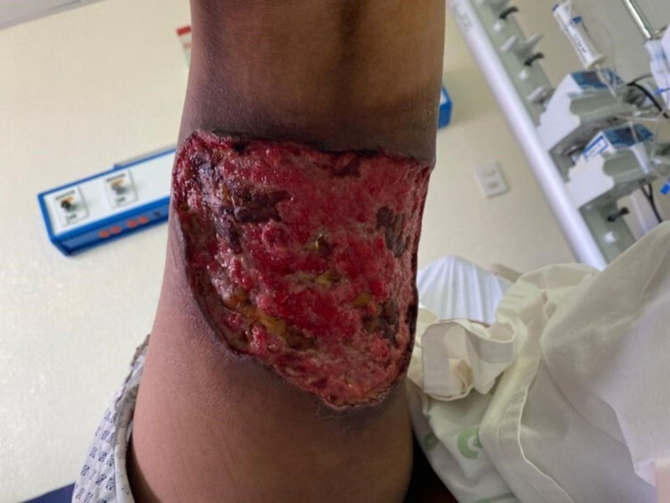
Figure 5. Lesion in the right pelvic limb after surgical lavage and debridement, with irregular edges, hyperpigmented, surrounding vitalized tissue.
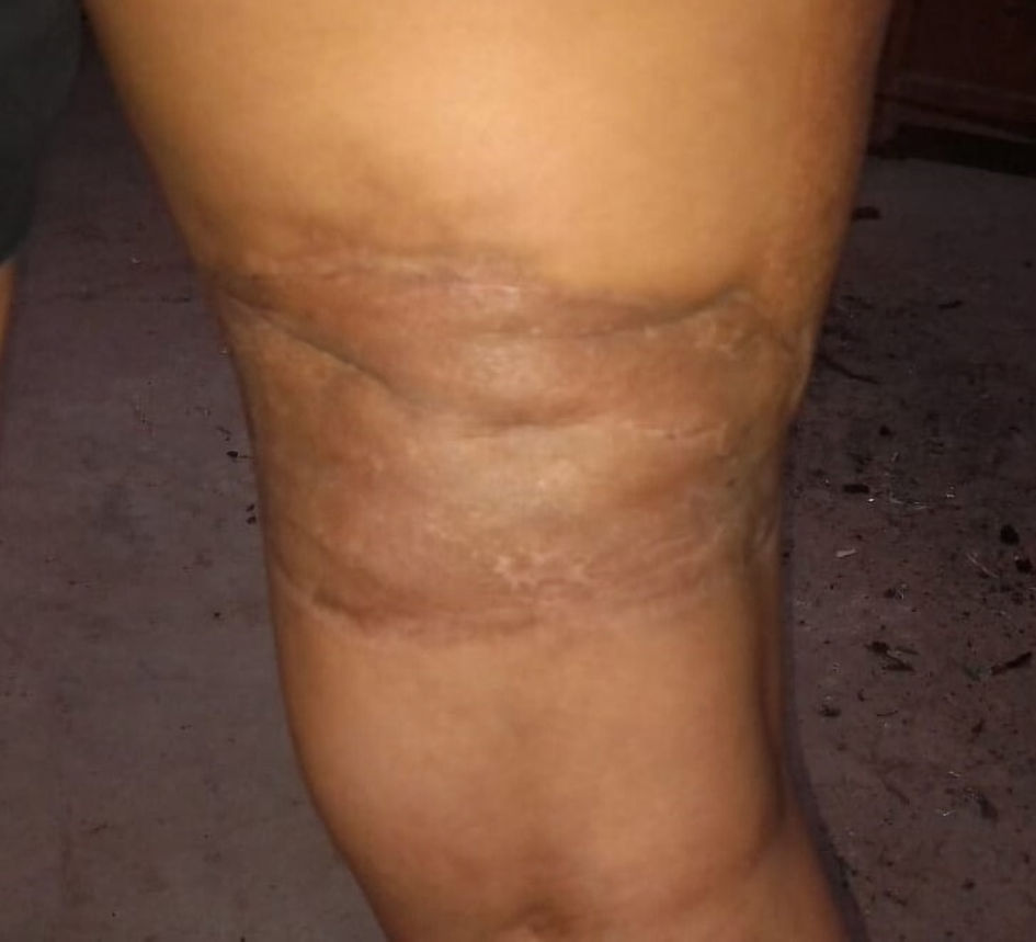
Figure 6. Complete resolution of the lesion 2 months after the attack, with residual scarring.
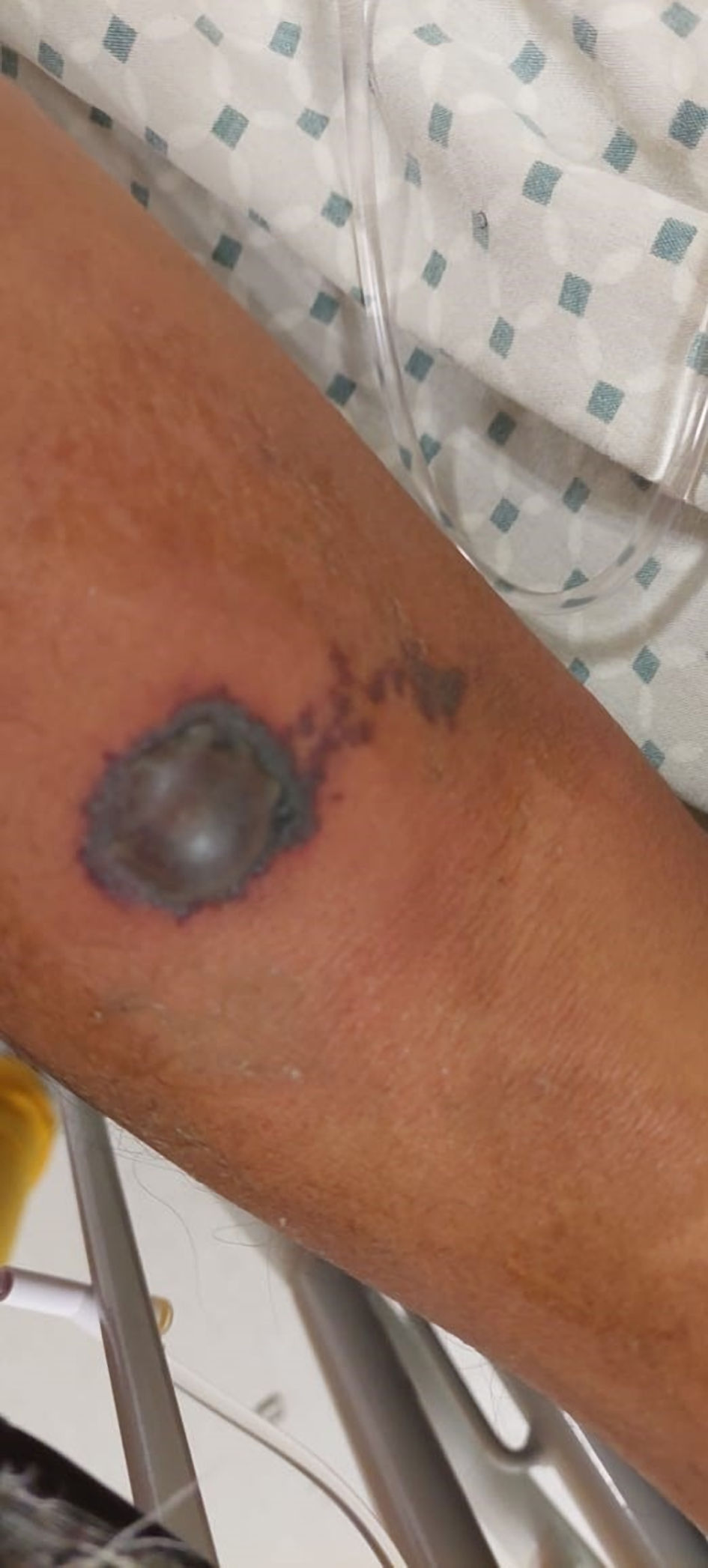
Figure 7. The skin lesion located on the right thoracic limb, with erythematous maculopapular characteristics, with serohematous content, irregular borders, with a white halo, together with a small diffuse liveloid plaque.
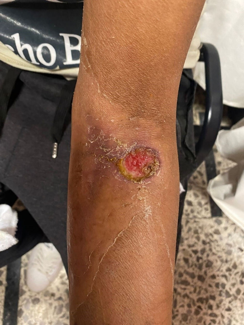
Figure 8. Ulcer with irregular borders, hyperpigmented, surrounded by vitalized tissue with fibrin formation after fabotherapeutic administration.
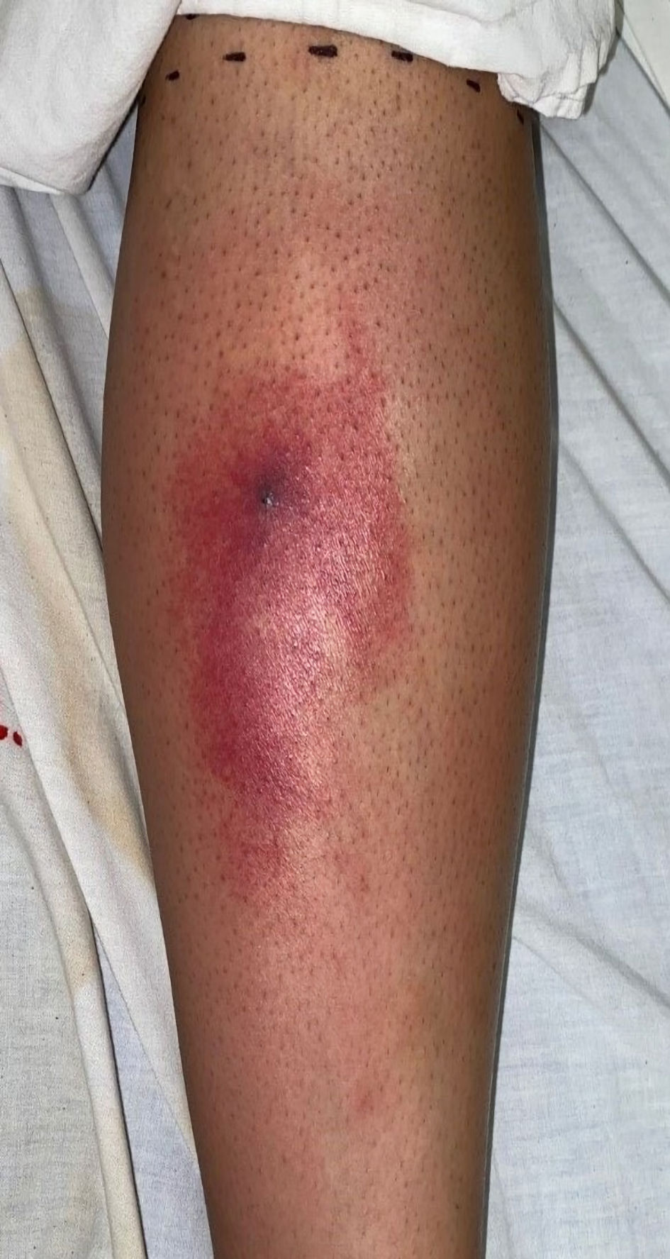
Figure 9. The lesion on the left lower extremity, in the area of the middle third of the medial tibia, of maculopapular type with a central necrotic area, with the presence of erythema with irregular borders.
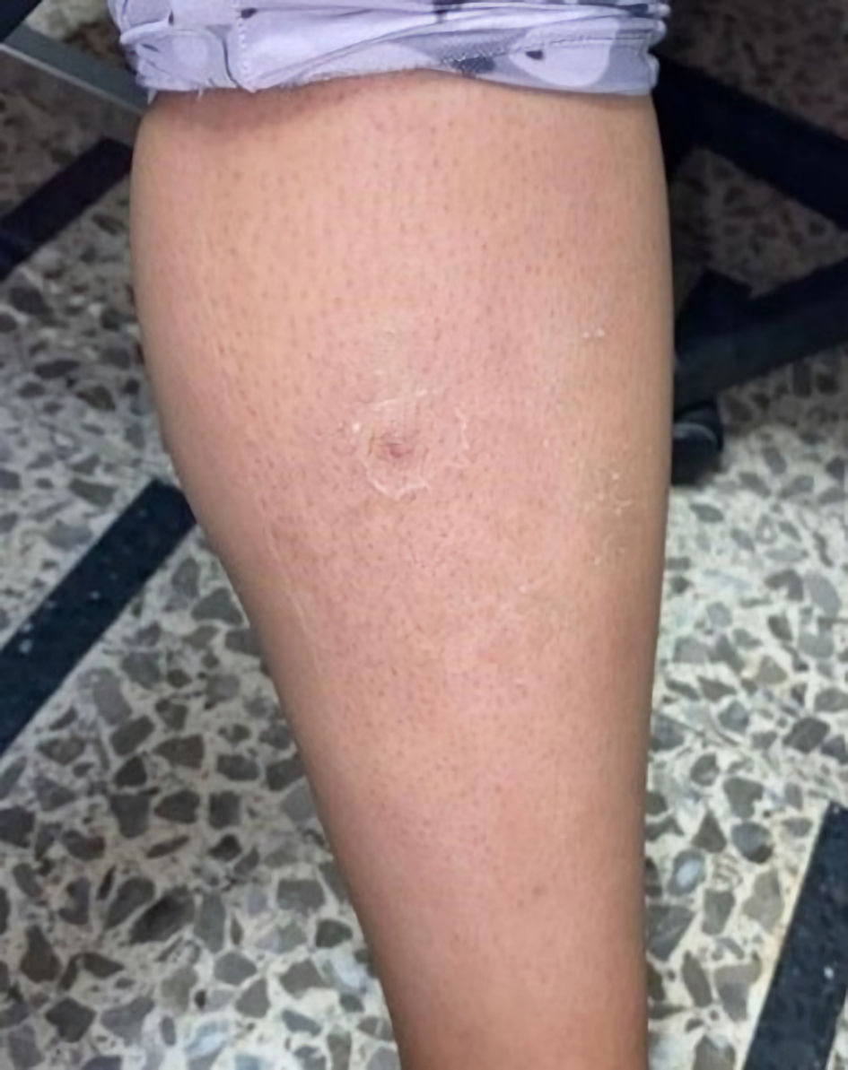
Figure 10. Complete resolution of the lesion 4 weeks after the attack, with residual scar.