Figures
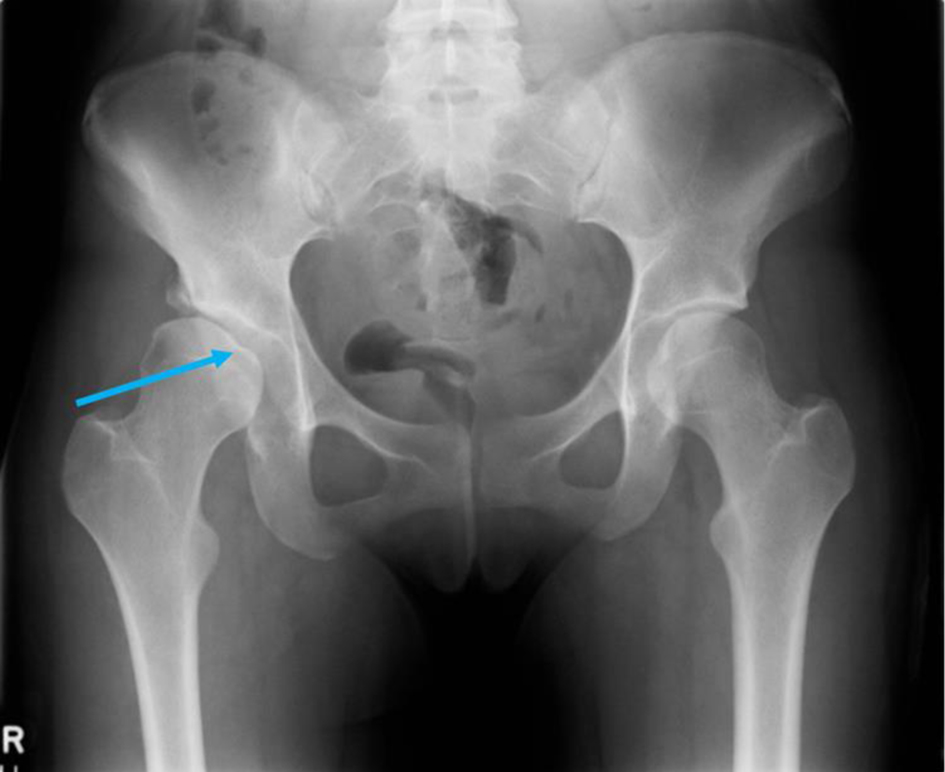
Figure 1. A plain radiograph taken during the first visit to the emergency department (ED), revealing right hip dysplasia, with uncovering of the lateral side of the femoral head and early secondary degenerative changes (blue arrow), and a normal appearance to the left hip on radiograph.
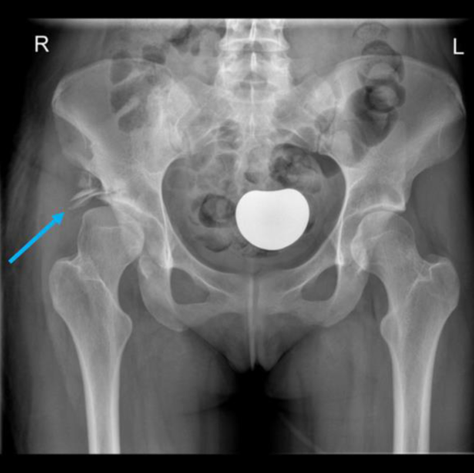
Figure 2. A plain radiograph after right labral repair with shelf osteotomy (blue arrow).
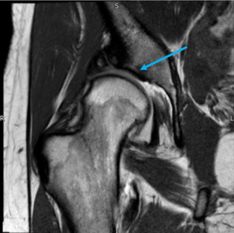
Figure 3. A magnetic resonance imaging (MRI) arthrogram of the right hip revealing a dysplastic hip and features of associated acetabular cartilage damage (blue arrow).
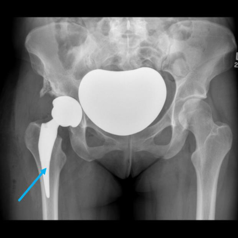
Figure 4. A plain radiograph showing a right total hip replacement (blue arrow).
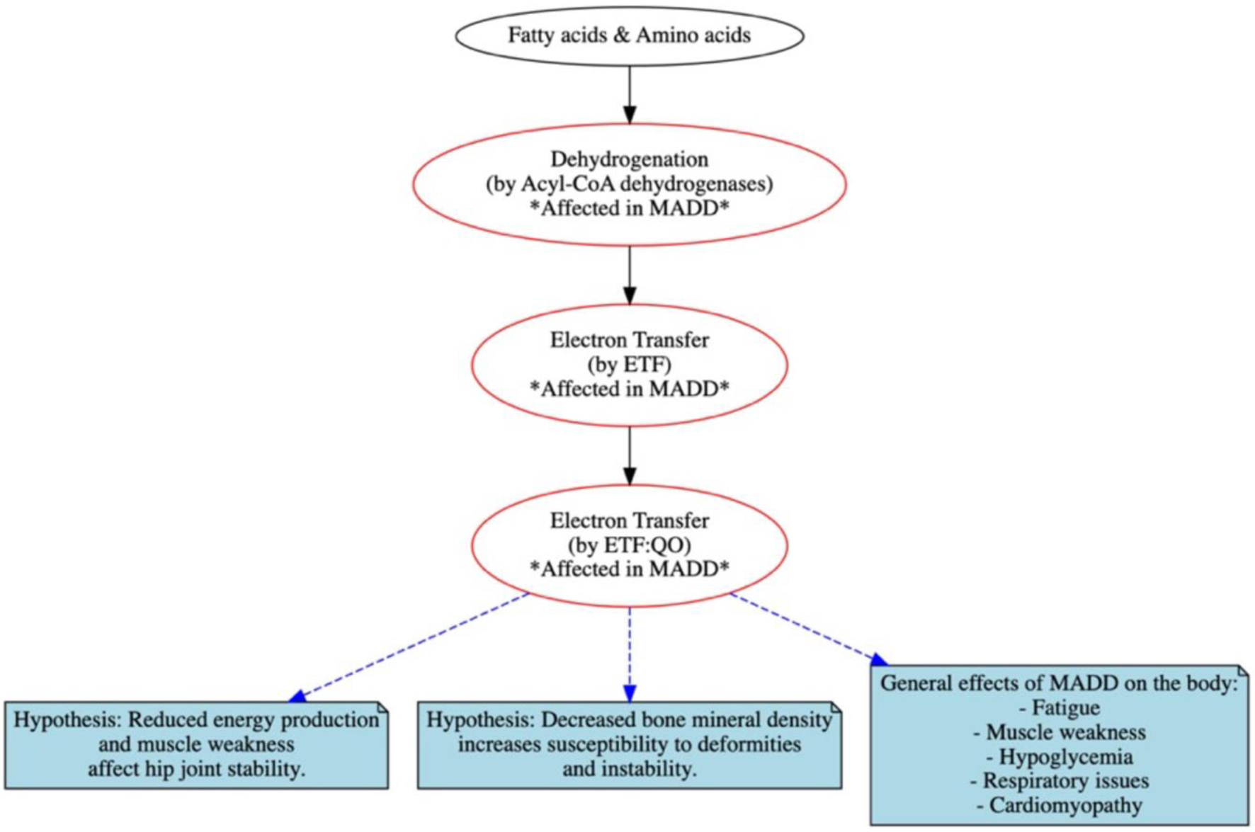
Figure 5. Schematic representation of key abnormalities including flavoprotein dehydrogenase enzymes, electron transfer flavoprotein (ETF) and ETF: ubiquinone oxidoreductase (ETF: QO) with outcomes in multiple acyl-CoA dehydrogenation deficiency (MADD).
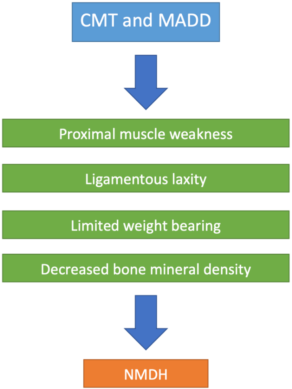
Figure 6. Suggested schematic representation of key abnormalities shared by Charcot-Marie-Tooth disease (CMT) and multiple acyl-CoA dehydrogenation deficiency (MADD) that in part can contribute to NMDH.