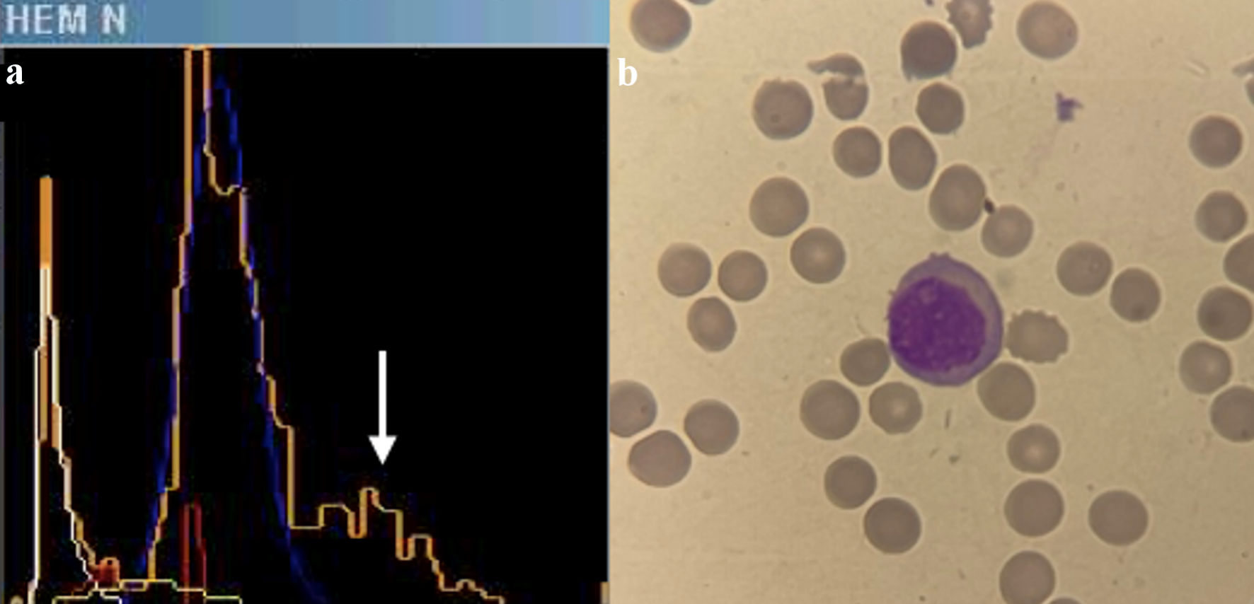
Figure 2. (a) Histogram of nucleated red blood cells in ADVIA 2021. A third peak is observed in orange (indicated by a white arrow), identifying non-stained cells with myeloperoxidase, which runs parallel to the lymphocyte peak in blue. (b) Plasma cell in peripheral blood film.
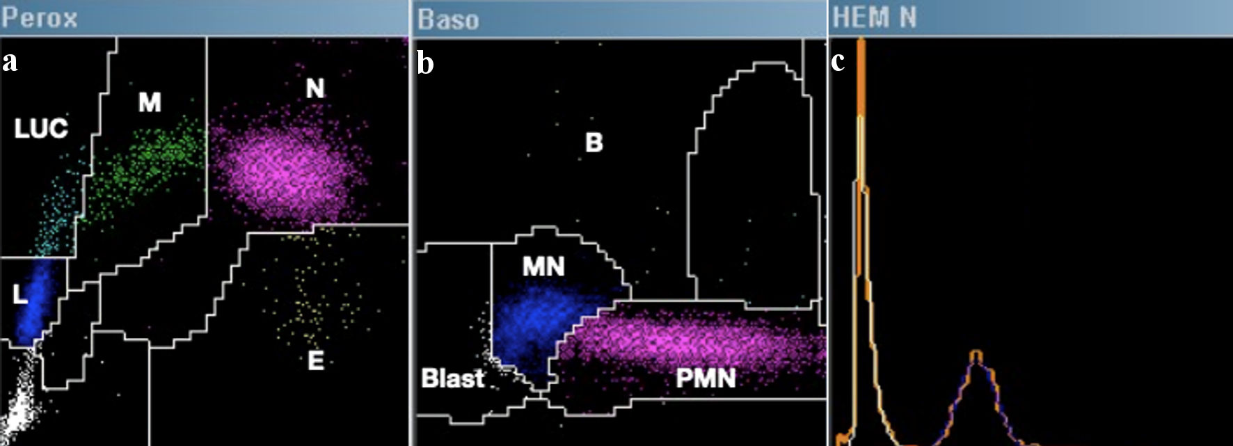
Figure 3. Example of a normal hematological count in ADVIA 2021. (a) Perox channel: E = eosinophils, N = neutrophils, M = monocytes, L = lymphocytes, LUCs = large unstained cells. (b) Baso channel: B = basophils, PMN = polymorphonuclear cells, MN: mononuclear cells, Blast = blasts. (c) The nucleated red blood cell enumeration histogram corresponds to the unstained area on the Y-axis of the Peroxidase channel. This visualization depicts overlaps of non-stained cells (orange), platelets (white), nucleated red blood cells if present (yellow and red), and lymphocytes (blue).