Journal of Medical Cases, ISSN 1923-4155 print, 1923-4163 online, Open Access
Article copyright, the authors; Journal compilation copyright, J Med Cases and Elmer Press Inc
Journal website https://www.journalmc.org

Case Report

Volume 15, Number 7, July 2024, pages 120-125


Organizing Pneumonia With Diffuse Alveolar Hemorrhage Induced by the Kampo Medicine Choreito

Figures

Figure 1.
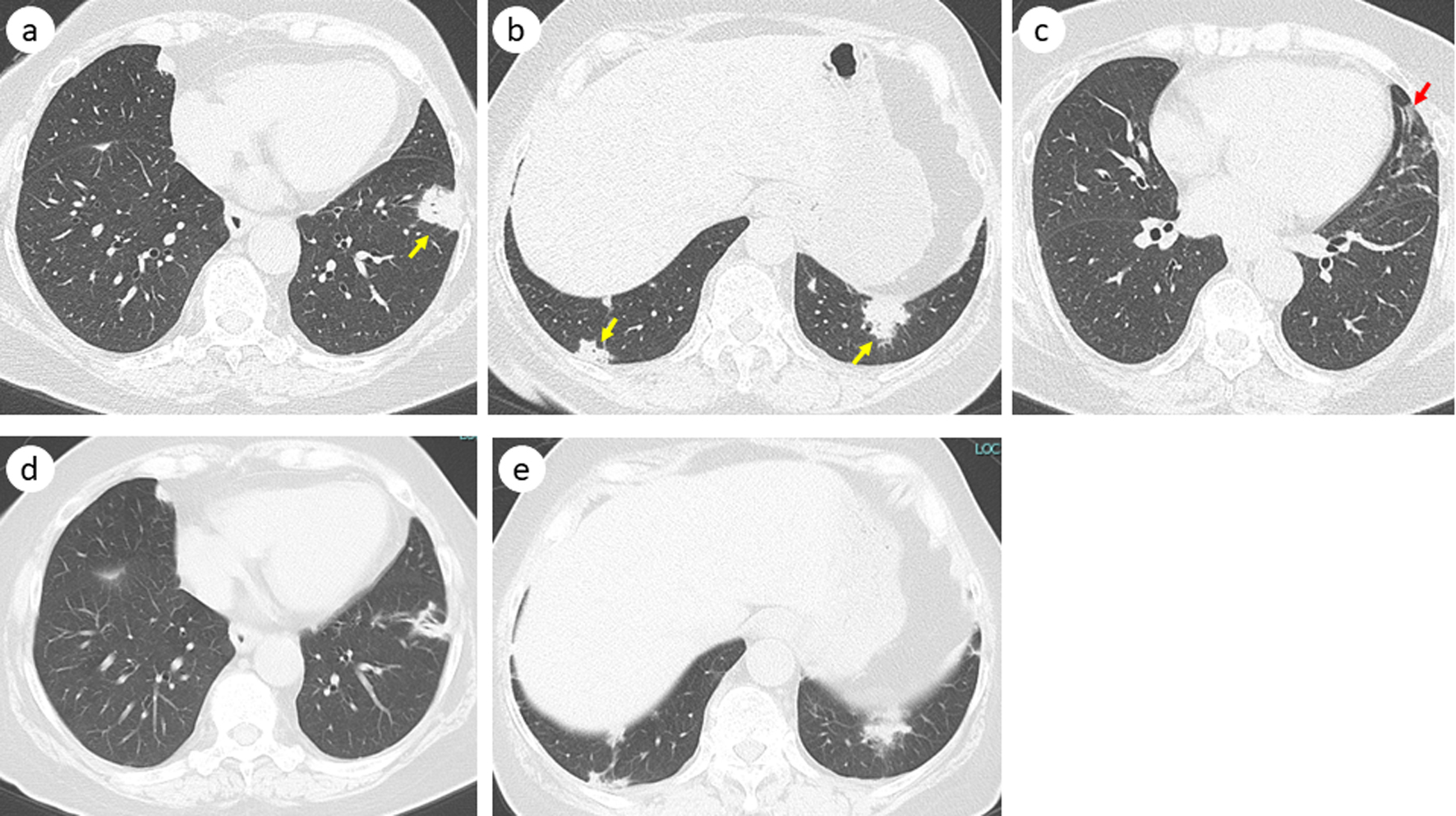
Figure 1. Chest computed tomography before bronchoscopy showing multiple nodules (yellow arrow) (a, b) and a slight ground-glass opacity in the left lingular lobe (red arrow) (c). Five months after discontinuation of choreito, these nodules shrank or vanished (d, e).
Figure 2.
Figure 2. Macroscopic findings of bronchoalveolar lavage fluid from the left B5. The numbers are the order of the samples obtained.
Figure 3.
Figure 3. Histology of bronchoscopic specimen from the tumor in the left B8a (hematoxylin and eosin staining, magnification × 400). The black arrows indicate an intraluminal polypoid structure with mild infiltration of chronic inflammatory cells.