Journal of Medical Cases, ISSN 1923-4155 print, 1923-4163 online, Open Access
Article copyright, the authors; Journal compilation copyright, J Med Cases and Elmer Press Inc
Journal website https://www.journalmc.org

Case Report

Volume 15, Number 7, July 2024, pages 126-129


An Atypical Presentation of Metastatic Renal Cell Carcinoma

Figures

Figure 1.
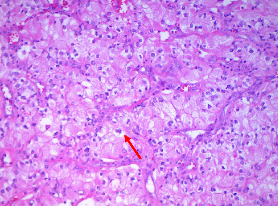
Figure 1. Surgical specimens from the sigmoid mass revealing neoplastic cells (arrow) with ample granular to clear cytoplasm, vesicular chromatin and scattered inflammatory cells growing in trabecular growth pattern (hematoxylin and eosin stain × 200).
Figure 2.
Figure 2. (a) CT angiography of the abdomen and pelvis showing a mass (arrow) in the splenic flexure of the colon. (b) Multiple hypodense lesions (arrowheads) in the liver suggestive of central necrosis. CT: computed tomography.

Table

Table 1. Notable Laboratory Values on Admission
 
Laboratory resultMeasurementReference
MCV: mean corpuscular volume; BUN: blood urea nitrogen; PTT: partial thromboplastin time; INR: international normalized ratio; TIBC: total iron-binding capacity.
Hemoglobin6.7 g/dL12.0 - 16.0 g/dL
MCV90.2 fL80.0 - 100.0 fL
Potassium5.2 mmol/L3.5 - 5.0 mmol/L
BUN30 mg/dL7 - 20 mg/dL
Albumin2.8 g/dL3.5 - 5.0 g/dL
PTT28 s25 - 35 s
INR1.090.9 - 1.1
Iron52 µg/dL60 - 170 µg/dL
TIBC249 µg/dL250 - 400 µg/dL
Percent saturation21%20 - 50%
Ferritin45 ng/mL15 - 200 ng/mL