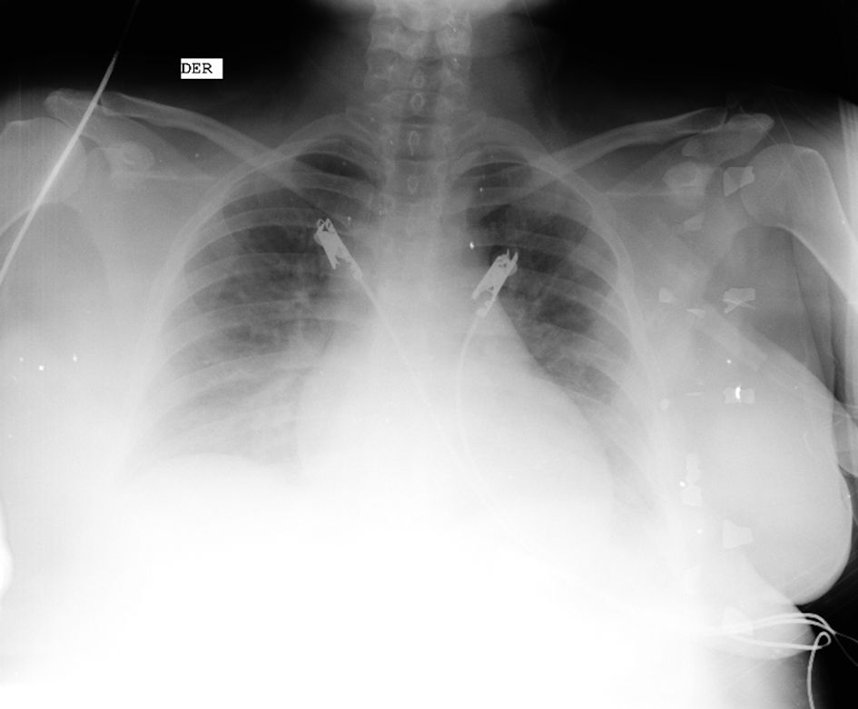
Figure 1. Chest X-ray. Figure shows enlarged cardiac silhouette, bilateral pulmonary vascular congestion and diffuse interstitial edema consistent with pulmonary congestion (source: patient’s medical chart, 2023).
| Journal of Medical Cases, ISSN 1923-4155 print, 1923-4163 online, Open Access |
| Article copyright, the authors; Journal compilation copyright, J Med Cases and Elmer Press Inc |
| Journal website https://www.journalmc.org |
Case Report
Volume 15, Number 8, August 2024, pages 171-179
Peripartum Cardiomyopathy: A Case Report of Mortality From a Rare and Potentially Fatal Condition
Figures
Tables
| Date | Time | Blood pressure (mm Hg) | Heart rate (beats/min) | Respiratory rate (breaths/min) | SaO2 (%) | Temperature (°C) |
|---|---|---|---|---|---|---|
| Source: patient’s medical chart, 2023. | ||||||
| August 22, 2023 | 12:50 | 82/60 | 112 | 22 | 99 | 37.8 |
| 16:19 | 109/64 | 120 | 19 | 96 | 36.8 | |
| 23:16 | 86/48 | 113 | 34 | 100 | 36.4 | |
| August 23, 2023 | 11:10 | 84/61 | 119 | 37 | 97 | 37.0 |
| 15:54 | 80/45 | 141 | 41 | 99 | 37.0 | |
| Date | pH | PO2 (mm Hg) | PCO2 (mm Hg) | HCO3 (mmol/L) | BE (mmol/L) | Lactate |
|---|---|---|---|---|---|---|
| Source: patient’s medical chart, 2023. PO2: partial pressure of oxygen; PCO2: partial pressure of carbon dioxide; HCO3: bicarbonate; BE: base excess. | ||||||
| August 22, 2023 | 7.47 | 104.0 | 25.0 | 18.2 | -4.1 | 2.4 |
| August 23, 2023 | 7.39 | 189.9 | 25.0 | 15.8 | -9 | 1.1 |
| Measurement | Patient values | Reference values |
|---|---|---|
| Source: patient’s medical chart, 2023. | ||
| Aortic root | 23 | 27 - 38 mm |
| Left atrium | 31 | 27 - 40 mm |
| Right ventricle | 23 | 21 - 35 mm |
| Septum in diastole | 11 | 6 - 11 mm |
| Left ventricle in diastole | 53 | 38 - 58 mm |
| Posterior wall in diastole | 11 | 6 - 11 mm |
| Left ventricle in systole | 48 | 21 - 40 mm |
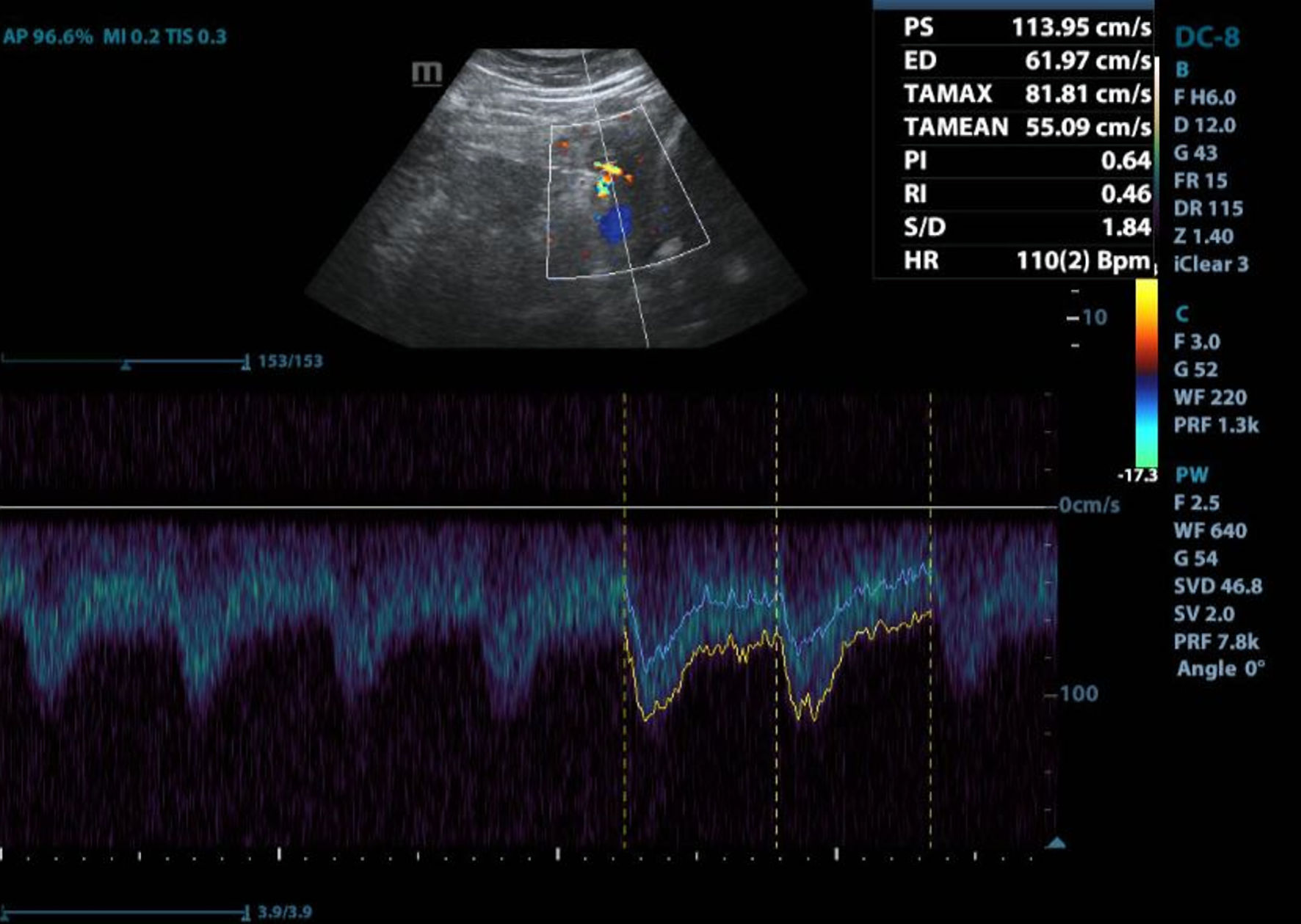
| Left ventricle ejection fraction | 23% | > 50% |