Journal of Medical Cases, ISSN 1923-4155 print, 1923-4163 online, Open Access
Article copyright, the authors; Journal compilation copyright, J Med Cases and Elmer Press Inc
Journal website https://www.journalmc.org

Case Report

Volume 15, Number 8, August 2024, pages 180-185


Primary Hyperparathyroidism and Pulmonary Embolism in Patients With a Fractured Neck of Femur

Figures

Figure 1.
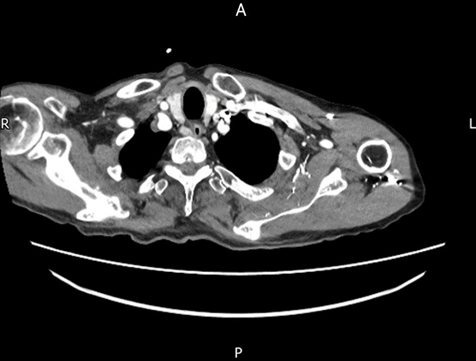
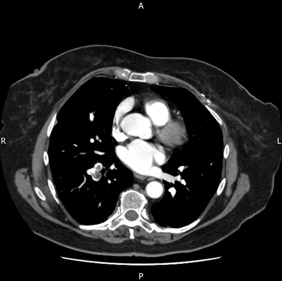
Figure 1. Axial view of computed tomography (CT) scan showing parathyroid nodule suspicious of adenoma.
Figure 2.
Figure 2. Sagittal view of computed tomography (CT) scan showing left intracapsular neck of femur fracture (yellow arrow).
Figure 3.
Figure 3. Axial view of computed tomography (CT) scan showing incidental finding of acute small volume PE in the right lobe (yellow arrow). PE: pulmonary embolism.
Figure 4.
Figure 4. Subcapital proximal femoral fracture of the neck of the femur (yellow arrow) in computed tomography (CT) scan.
Figure 5.
Figure 5. MRI confirming fracture diagnosis of right neck of femur with no lytic lesions. MRI: magnetic resonance imaging.
Figure 6.
Figure 6. A CT chest/abdomen/pelvis scan was performed, revealing bilateral lower lobe pulmonary emboli without evidence of malignancy. CT: computed tomography.
Figure 7.
Figure 7. The potential interplay between bone fracture, hypercalcemia, comorbidities, and high PTH. DVT: deep vein thrombosis; PE: pulmonary embolism; HPT: hyperparathyroidism: PLT: platelets; NOF: fracture neck of femur; PTH: parathyroid hormone.

Table

Table 1. PTH, Adjusted Calcium, Calcium-Creatinine Ratio and Vitamin D Levels for Both Cases
 
ParameterCase 1 (82 years old)Case 2 (77 years old)Reference range
PTH: parathyroid hormone.
PTH15.29.21.6 - 6.9 pmol/L
Adjusted calcium2.93.052.1 - 2.6 mmol/L
Urine calcium/creatinine ratio0.390.420.07 - 0.28
Vitamin D50.35550.0 - 150.0 nmol/L