Figures
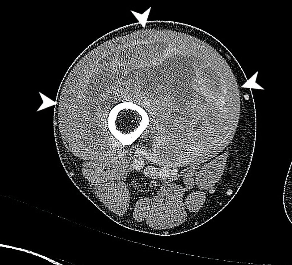
Figure 1. CT axial imaging of the right thigh at the time of initial presentation demonstrates a heterogeneously enhancing mass deep to vastus musculature (arrowheads). This circumferentially surrounds the anterior femur with cortical scalloping and irregularity at anterior aspect. The common femoral artery and vein, superficial femoral artery and vein and sciatic nerve, are all free of tumor (all boundaries are not fully delineated in this single slice). CT: computed tomography.
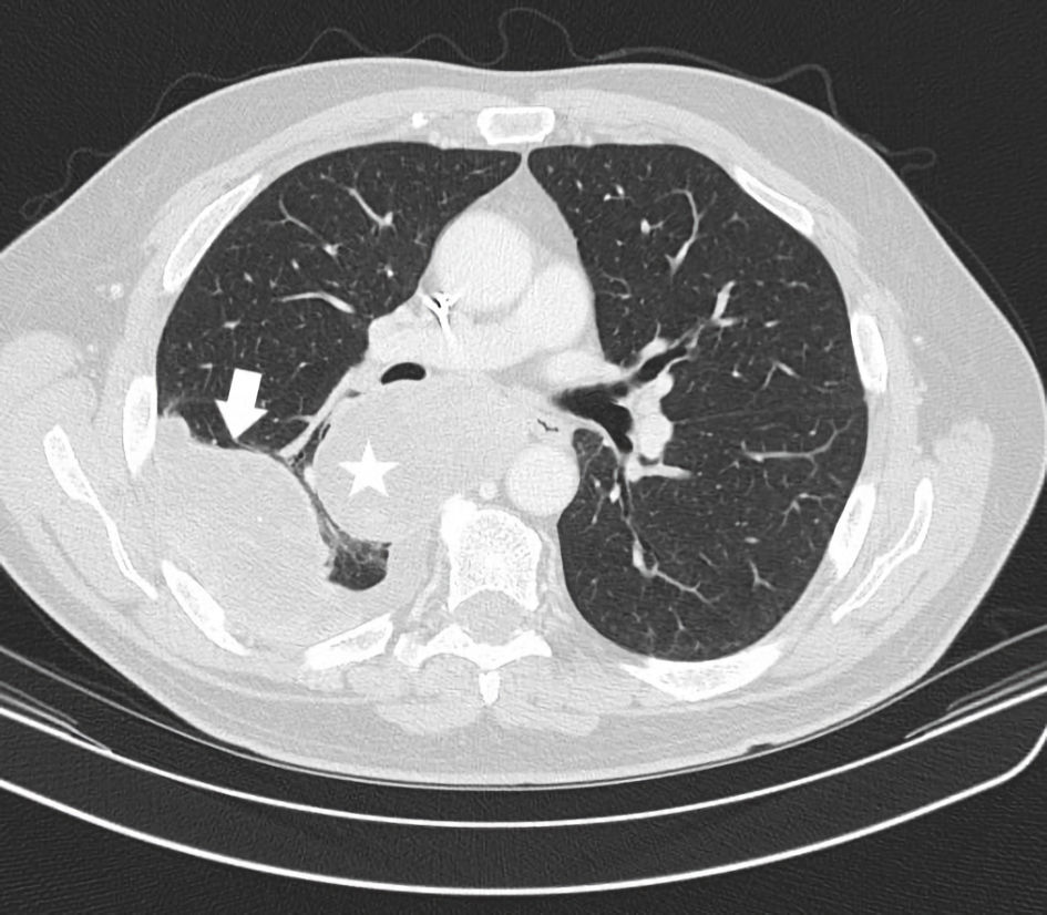
Figure 2. CT axial imaging of chest just prior to instituting immunotherapy shows the status of prominent pulmonary metastases. A right perihilar mass posterior to the right mainstem bronchus (star) and a separate right lower lobe pleural based mass with small effusion (arrow) are depicted. CT: computed tomography.
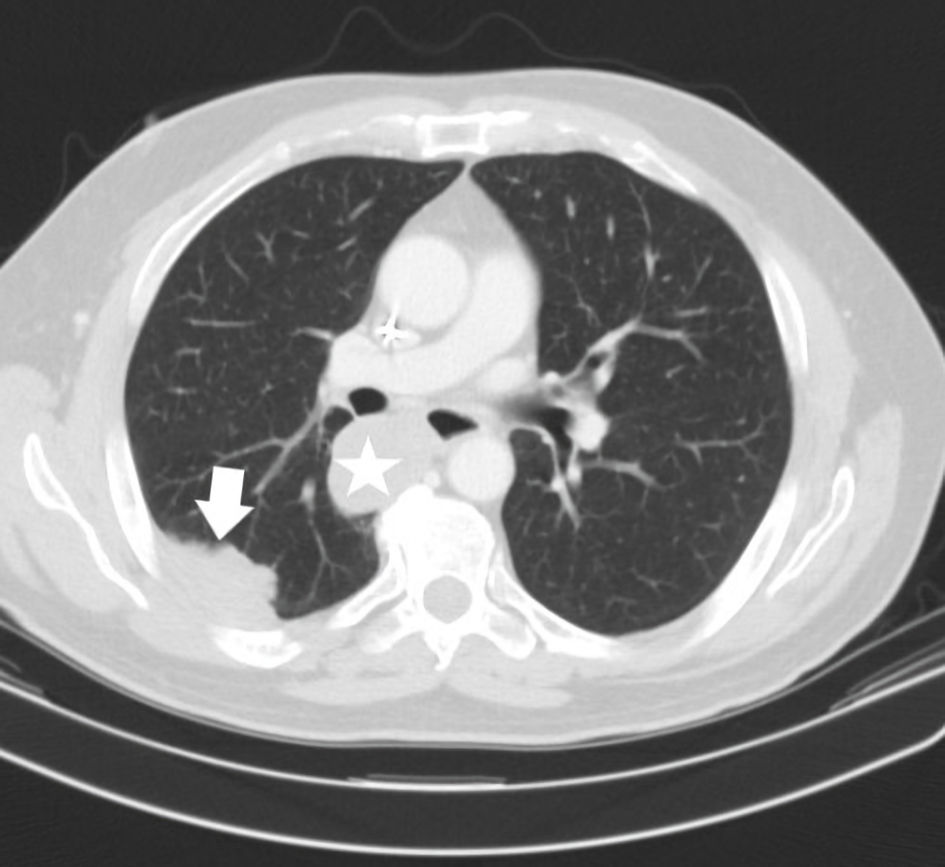
Figure 3. CT axial imaging of chest (January 2018) was performed as initial restaging to assess an early immunotherapy response. This demonstrates a substantially reduced size of the right perihilar mass now measuring 4.5 cm, previously 7.4 cm (star). The right lower lobe pleural mass has reduced to 5.2 cm, previously 7.4 cm (arrow). CT: computed tomography.
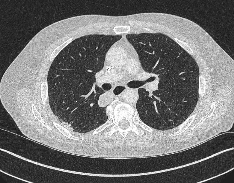
Figure 4. CT axial imaging of chest (October 2019) after the patient received immunotherapy for 2 years, demonstrates resolution of the right lower lobe pleural/subpleural mass (compare with Figure 3) and a decreased right perihilar mass measuring 3 cm (not depicted in same image due to different axial slice). CT: computed tomography.
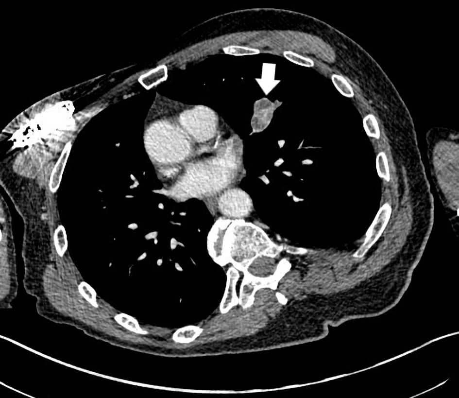
Figure 5. CT axial imaging of the chest indicates a left upper lobe mass measuring 3.4 ×2.5 cm (arrow) which became unresponsive to immunotherapy and progressed. CT: computed tomography.
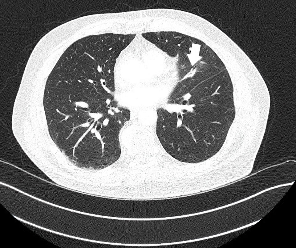
Figure 6. CT axial imaging of the chest 2 months post-irradiation with continued immunotherapy shows improved size of left upper lobe mass now 1.4 × 0.8 cm (arrow). This lesion disappeared with successive restaging by the following year with ongoing immunotherapy. CT: computed tomography.