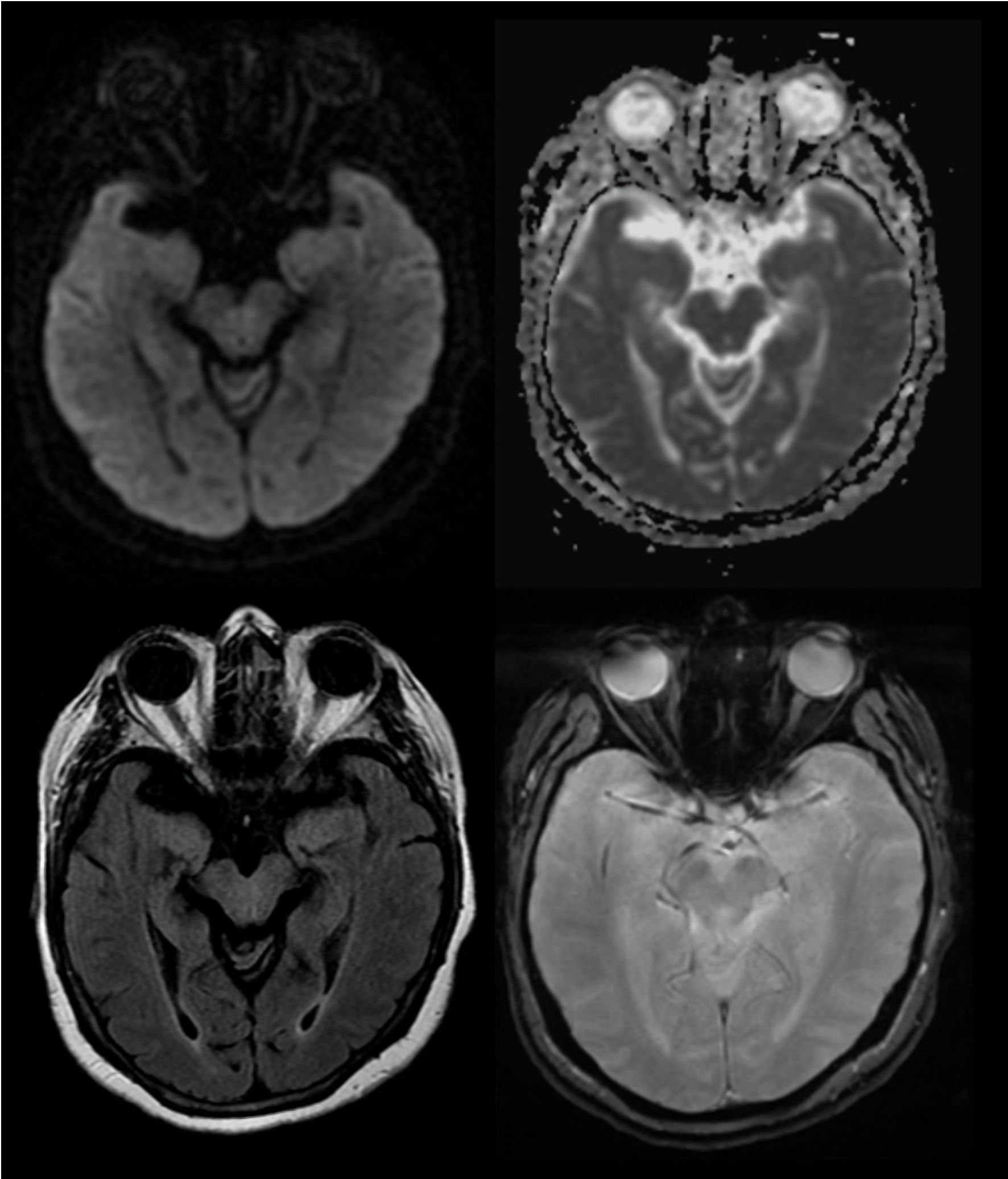
Figure 1. MRI upon presentation. The MRI shows no evidence of acute ischemic stroke on DWI (top left) or FLAIR lesion (bottom left). Pictured in the top right is the ADC and the bottom right is the GRE. MRI: magnetic resonance imaging; DWI: diffusion-weighted imaging; FLAIR: fluid-attenuated inversion recovery; ADC: apparent diffusion coefficient; GRE: gradient echo sequence.
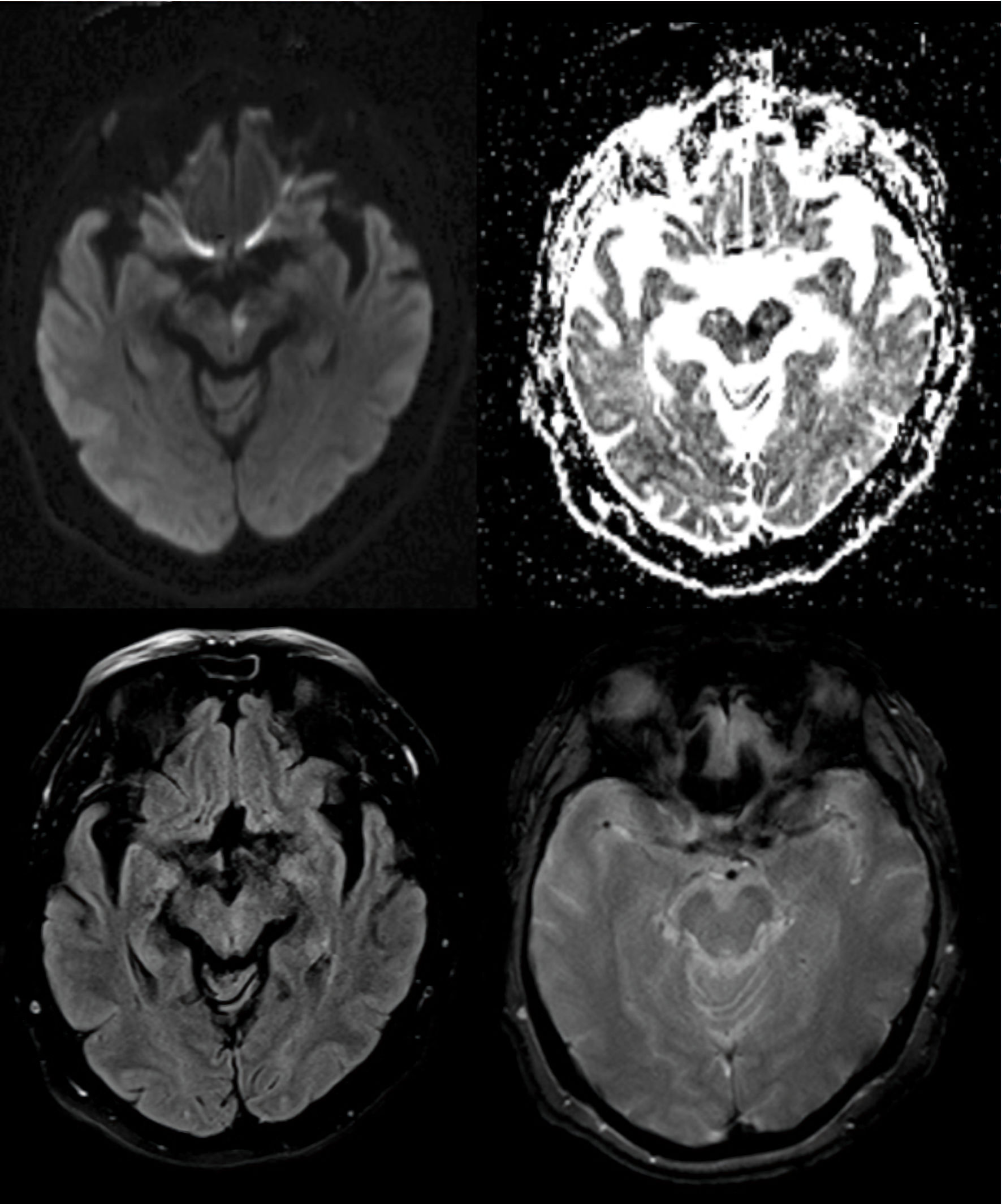
Figure 2. MRI 48 h post presentation. Abnormal MRI now shows a left MLF lesion on the DWI (top left) with an ADC correlate (top right) as well as a faint FLAIR abnormality (bottom left). Absence of any post thrombolytic hemorrhage is noted on the GRE (bottom right). MRI: magnetic resonance imaging; MLF: medial longitudinal fasciculus; DWI: diffusion-weighted imaging; FLAIR: fluid-attenuated inversion recovery; ADC: apparent diffusion coefficient; GRE: gradient echo sequence.