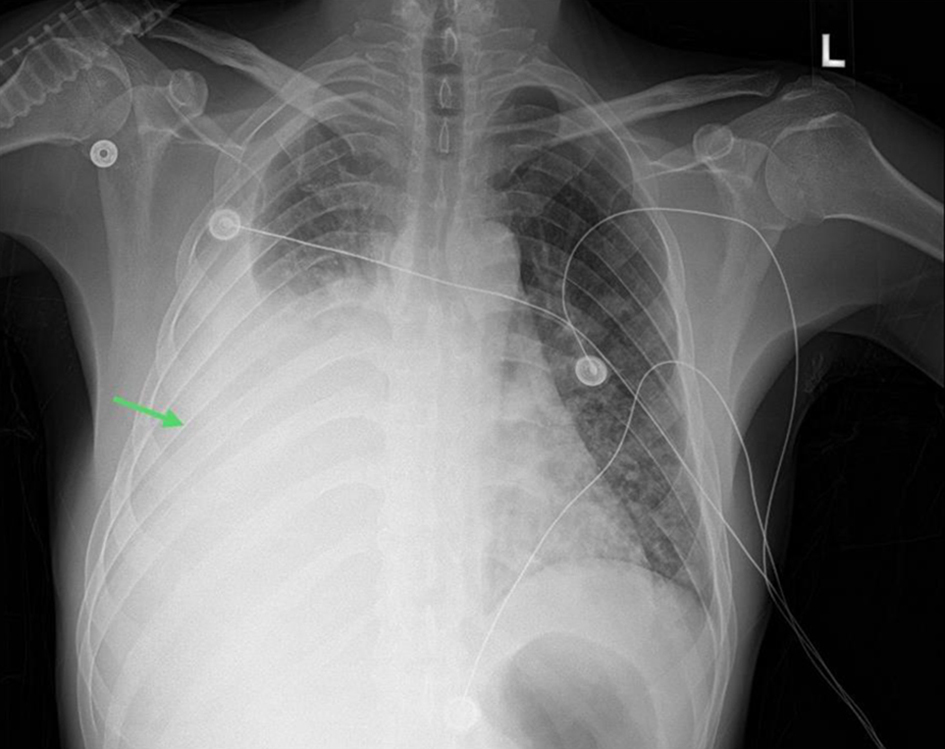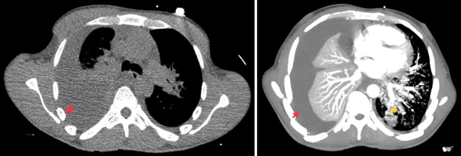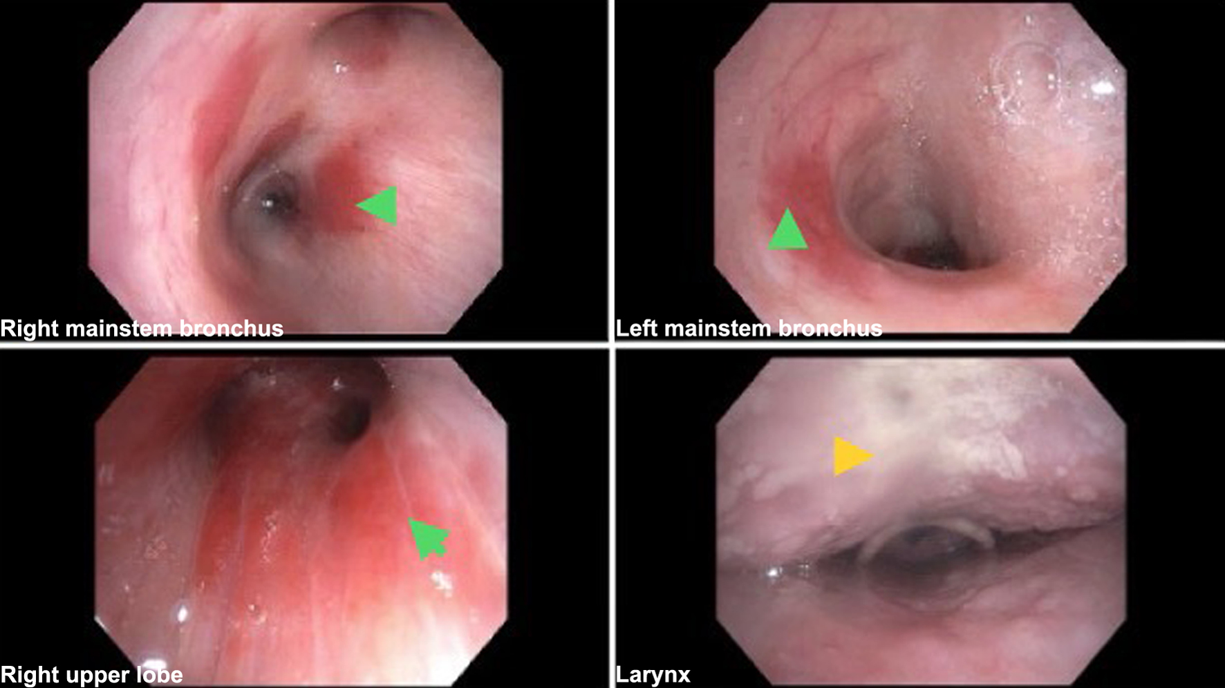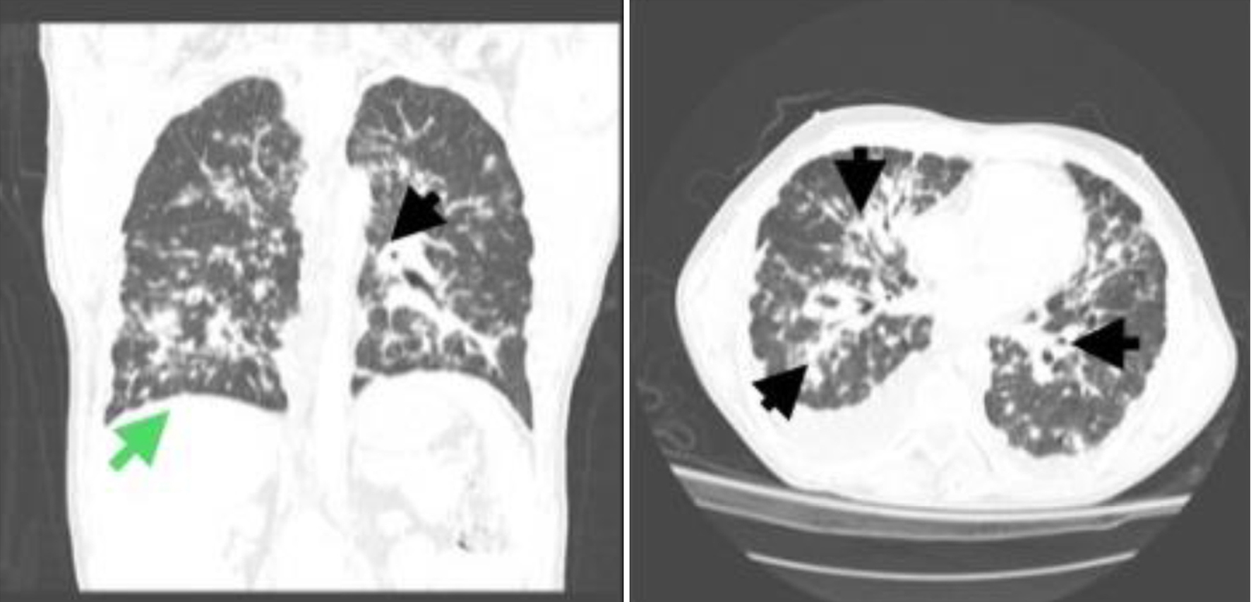
Figure 1. Frontal view chest X-ray showing large right-sided pleural effusion with adjacent atelectasis (green arrow).
| Journal of Medical Cases, ISSN 1923-4155 print, 1923-4163 online, Open Access |
| Article copyright, the authors; Journal compilation copyright, J Med Cases and Elmer Press Inc |
| Journal website https://www.journalmc.org |
Case Report
Volume 15, Number 11, November 2024, pages 311-318
Pulmonary Kaposi Sarcoma in the Era of Antiretroviral Therapy: A Case Series
Figures


Tables
| Laboratory test | Case 1 | Case 2 | Reference value |
|---|---|---|---|
| ALT: alanine transaminase; AST: aspartate aminotransferase; eGFR: estimated glomerular filtration rate; FTA: fluorescent treponemal antibody; HIV-1: human immunodeficiency virus 1; qPCR: quantitative polymerase chain reaction; RPR: rapid plasma reagin; STD: sexually transmitted disease. | |||
| Sodium | 122 | 128 | 135 - 145 mEq/L |
| Serum albumin | 3.2 | 4.0 | 3.5 - 5.7 g/dL |
| Total protein | 7.4 | 10.3 | 6.4 - 8.4 g/dL |
| Blood urea nitrogen | 9 | 15 | 7 - 23 mg/dL |
| Creatinine | 0.42 | 0.93 | 0.60 - 1.30 mg/dL |
| eGFR | 146 | 104 | ≥ 60 mL/min/1.73 m2 |
| Alkaline phosphatase | 108 | 157 | 34 - 104 U/L |
| AST | 25 | 92 | 13 - 39 U/L |
| ALT | 13 | 78 | 7 - 52 U/L |
| Troponin level | < 2 | 3 | 3 - 17 pg/mL |
| B-type natriuretic peptide level | 29 | 9 | 1 - 100 pg/mL |
| White blood cell count | 2.6 | 7.6 | 4.5 - 11.0 × 10³/mm3 |
| Hemoglobin level | 8.9 | 10.1 | 12.0 - 16.0 g/dL |
| Platelet count | 272 | 170 | 140 - 440 × 103/mm3 |
| D-dimer | 7.18 | ≤ 0.50 | |
| HIV-1 RNA viral load | 134,000 | 2,860,000 | |
| Absolute CD4 helper count | 93 | 163 | 359 - 1,519 cells/mm3 |
| Pneumocystis carinii pneumonia qPCR | Negative | Negative | |
| Lactate dehydrogenase level | 135 | 233 | 140 - 271 U/L |
| Ferritin level | 596.0 | 57 | 16.4 - 294 ng/mL |
| C-reactive protein | 6.8 | ≤ 9.9 mg/dL | |
| Erythrocyte sedimentation rate | 83 | 0 - 10 mm/h | |
| Lactic acid | 1.0 | 0.5 - 2.2 mmol/L | |
| FTA for Treponema pallidum | Reactive | Negative | |
| Treponema pallidum antibodies | Reactive | Negative | |
| RPR titer | 1:1 | ||
| STD panel | |||
| C3 and C4 complement | Normal | ||
| Parameter | Case 1 | Case 2 |
|---|---|---|
| LDH: lactate dehydrogenase; RBC: red blood cell; WBC: white blood cell. | ||
| Specific gravity | 1.346 | 1.360 |
| Urea | 9 mg/dL | |
| Color | Orange | Bloody |
| Appearance | Cloudy | Cloudy |
| WBC | 443/mm3 | 1,400/mm3 |
| RBC | 48,000/mm3 | 100,000/mm3 |
| Neutrophil | 4% | 4% |
| Lymphocyte | 63% | 71% |
| Monocytes | 21% | 11% |
| Histocyte | 10% | 2% |
| Mesothelial cell | 2% | 12% |
| Albumin | 2.7 g/dL | 2.2 g/dL |
| Glucose | 105 mg/dL | 80 mg/dL |
| LDH | 136 U/L | 967 U/L |
| Protein | 5.4 g/dL | 6.8 g/dL |
| Cholesterol | 53 mg/dL | |
| Triglycerides | 35 mg/dL | |