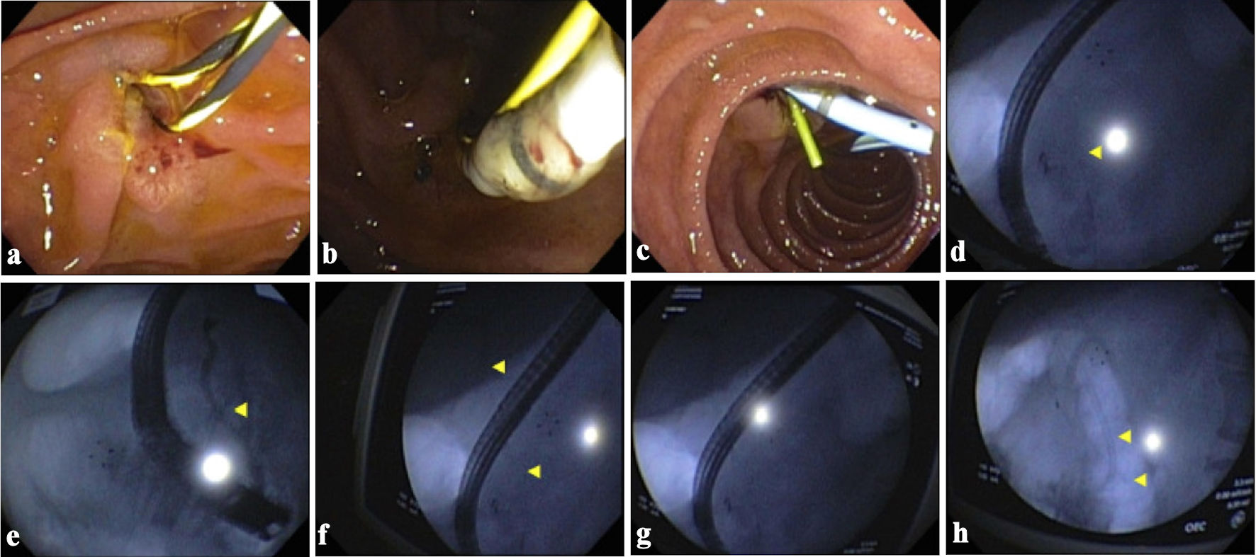
Figure 1. Endoscopic retrograde cholangiopancreatography images showing a filling defect in the biliary duct, sphincterotomy, and stent placement. (a) Image showing a short 0.035-inch soft Jagwire being passed into the biliary tree. (b) Endoscopic image showing a cannulating sphincterotome being inserted into the biliary tree. (c) Image showing a one 4-Fr 5-cm plastic stent with a single external flap and no internal flaps being placed into the common bile duct (CBD)/ventral pancreatic duct (PD). (d) CBD wire. (e) PD w/cutoff a level of genu. (f-h) CBD wire. Yellow arrows in (d-f) show a filling defect consistent with a stone in genu of the pancreas. Yellow arrows in (f) and (h) show a filling defect consistent with a stone as seen on the cholangiogram. w/cutoff: with cutoff.
