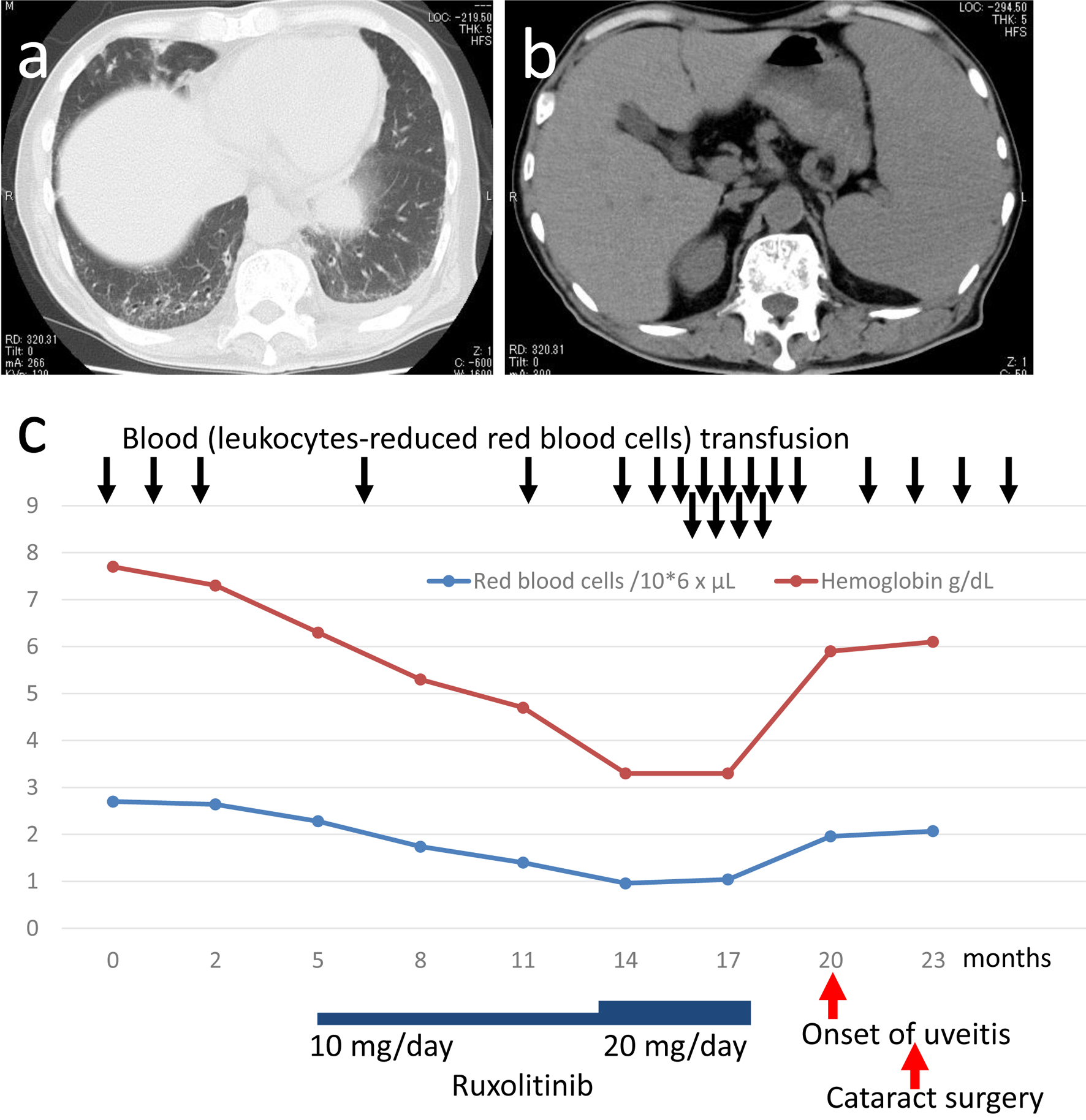
Figure 1. Computed tomographic scans (a, b) at the initial visit and a chart for the entire course (c). Reticular and ground glass appearance, indicative of interstitial change, in bilateral lower dorsal lung fields with a small amount of pleural effusion on the left side (a). Hepatosplenomegaly (b).

Figure 2. Bone marrow biopsy to support the diagnosis of myelofibrosis. Conspicuous fibrosis with osteosclerosis and reduced amount of adipose tissue in low magnification (a), and cluster of megakaryocytes with bizarre nuclei (arrow, b) in high magnification by hematoxylin-eosin stain. Silver impregnation stain reveals diffusely increased reticulin fibers (c). Masson trichrome stain highlights collagen fibrosis (d). By immunostaining, clusters of CD42b-positive bizarre megakaryocytes (e), CD71-positive erythroid lineage cells in erythroid islands which are in reduced number and in smaller size (f), and myeloperoxidase (MPO)-positive myeloid lineage cells in the normal process of maturation (g). Scale bar = 500 µm in A, 50 µm in B, 200 µm in c-g.
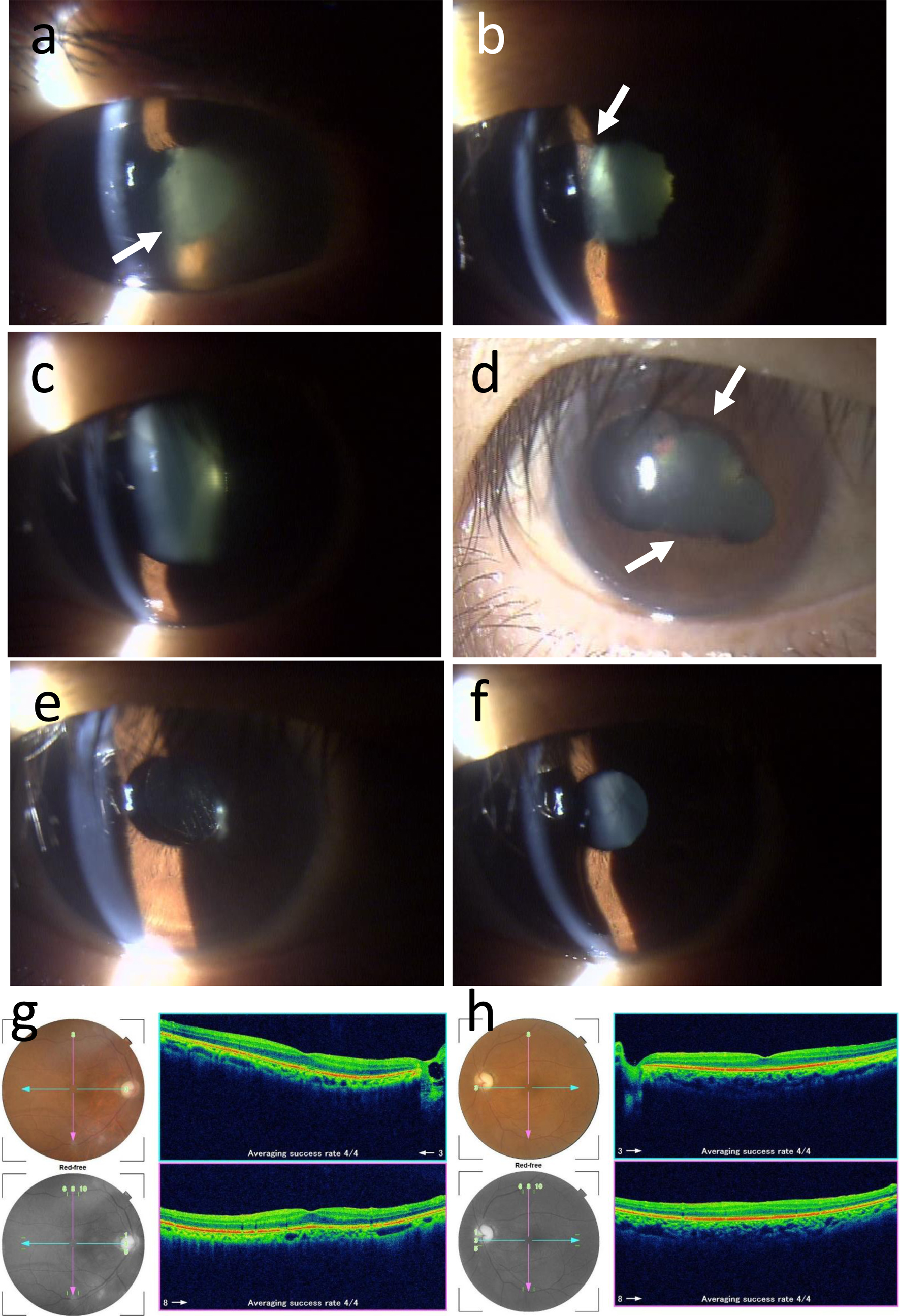
Figure 3. Slit-lamp photographs at ophthalmic presentation (right eye, a), a week later (right eye, b), 3 weeks later (right eye, c, d), 2 months later (right eye, e; left eye, f) from the first ophthalmic visit. Fundus photographs and optical coherence tomography (g, h) 2 months later. Fluffy fibrin deposition in the pupillary area (arrow in a) of anterior chamber has disappeared a week later (b) only with topical 0.1% betamethasone eye drops six times daily, leaving iris posterior synechia (arrow in b). Upper and lower iris posterior synechia (arrows in d) under mydriasis with 1% atropine eye drops 3 weeks later (c, d). No inflammation after intraocular lens implantation by cataract surgery in the right eye (e), and also in the left eye with anterior and posterior subcapsular cataract (f). Ocular fundus photographs and optical coherence tomographic images in the right eye (g) and left eye (h) appear normal except for temporal pallor of the optic disc in the right eye and total atrophy of the optic disc in the left eye. Each panel in g and h shows color fundus photograph (top left), red-free photograph (bottom left), horizontal section (shown in blue arrows in photographs) of the image from the nasal to the temporal side (top right), and vertical section (shown in pink arrows in photographs) of the image from the superior to the inferior side (bottom right).