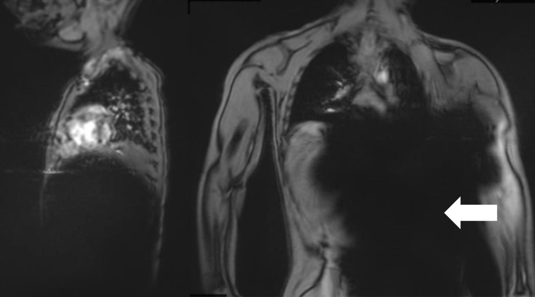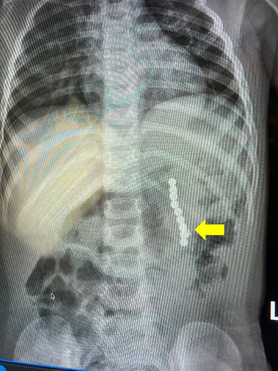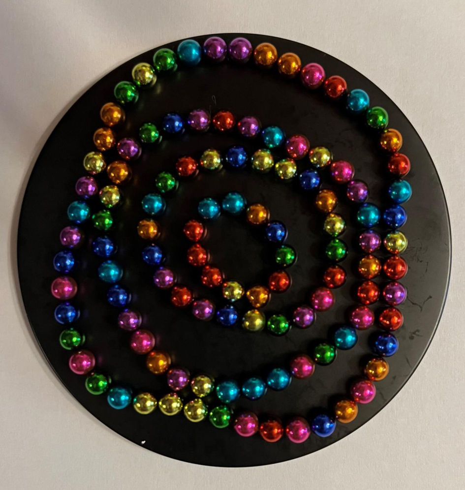
Figure 1. Initial magnetic resonance imaging scout sequences demonstrating prominent susceptibility artifacts (white arrow) emanating from the abdomen suggestive of a ferromagnetic foreign body.
| Journal of Medical Cases, ISSN 1923-4155 print, 1923-4163 online, Open Access |
| Article copyright, the authors; Journal compilation copyright, J Med Cases and Elmer Press Inc |
| Journal website https://www.journalmc.org |
Case Report
Volume 15, Number 11, November 2024, pages 319-323
Ingested Magnets Found Inadvertently During Elective Magnetic Resonance Imaging
Figures


Table
| Author and reference | Patient demographics | Summary and outcome |
|---|---|---|
| CT: computed tomography; ED: emergency department; MRI: magnetic resonance imaging; POD: postoperative day. | ||
| Bailey et al [8] | A 5-year-old male who presented to the ED with complaint of neck pain and refusal to move his neck. | MRI of the brain and cervical spinal cord were normal. The patient was kept overnight for observation and pain control. The following day, the patient developed severe abdominal pain and refused to eat. Radiographs of the abdomen revealed pneumoperitoneum and 11 small round objects in the left upper quadrant. Exploratory laparotomy revealed four 5 - 7 mm full thickness small bowel perforations and 11 small, round magnets. The patient was discharged home on POD 8. |
| Metterlein et al [9] | A 3-year-old male who presented to the ED with a 10-day history of an upper respiratory infection that had progressed to include diffuse swelling and redness of the forehead and the right orbital region. | MRI of the head was obtained to plan a possible surgical intervention. Extinction of facial imaging due to a ferromagnetic object in the nasal region was noted. The scan was terminated, and manual inspection of the nasal cavity revealed a small button battery. There was generalized swelling and a small mucosa lesion consistent with a burn or acid corrosion. The patient was discharged home after 3 days. |
| Glover et al [10] | A 65-year-old man with schizophrenia and intellectual disability who presented to the ED with abdominal pain. | MRI of the head was performed the previous day, but stopped due to abdominal complaints. The patient was sent home, but presented to the ED the following day with pain and systemic signs of infection. CT imaging revealed a metallic artifact and pneumoperitoneum. Exploratory laparotomy revealed two 3.5 cm metal sockets and a clevis pin. There was a serosal defect in the stomach. The patient was discharged home on POD 14. |