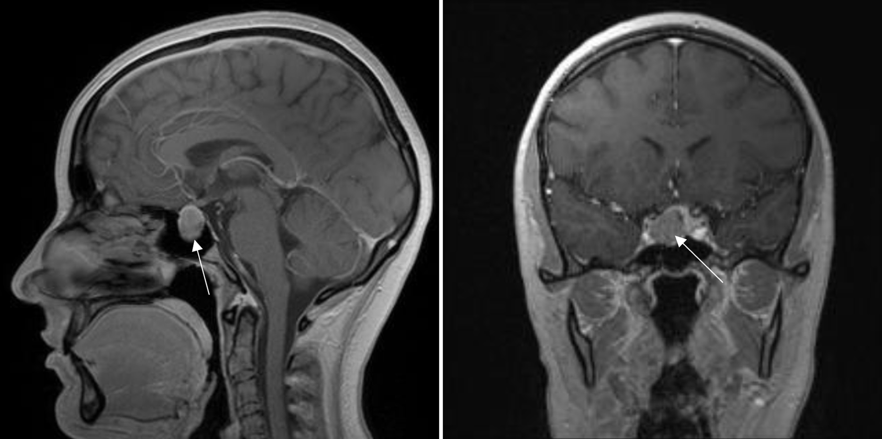
Figure 1. Magnetic resonance imaging examination showing hypophysial prolactinoma, suprasellar formation till optic chiasma (arrow).
| Journal of Medical Cases, ISSN 1923-4155 print, 1923-4163 online, Open Access |
| Article copyright, the authors; Journal compilation copyright, J Med Cases and Elmer Press Inc |
| Journal website https://www.journalmc.org |
Case Report
Volume 15, Number 9, September 2024, pages 242-249
Intraoperative Takotsubo Syndrome
Figures