Figures

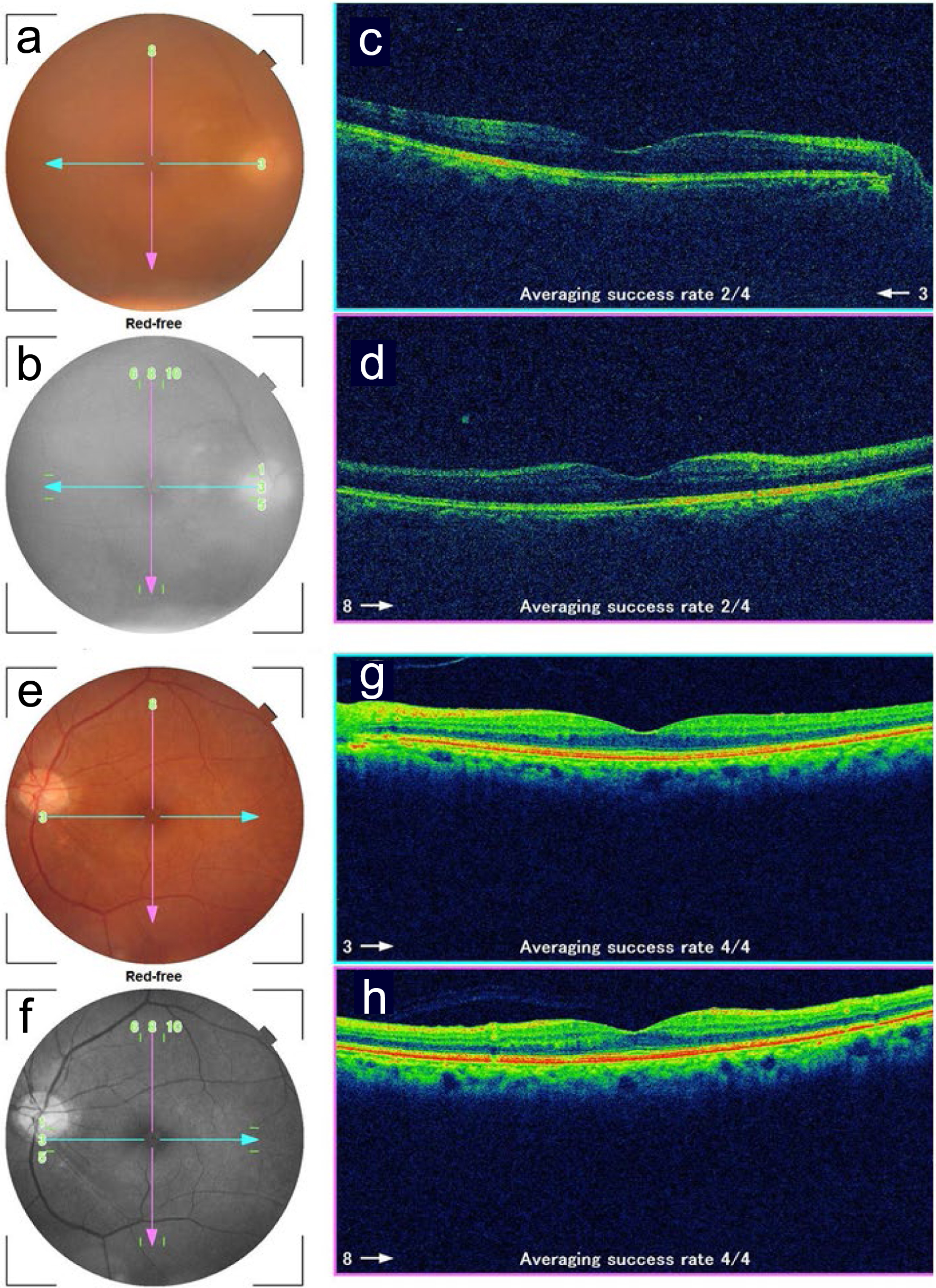
Figure 1. Fundus photographs and optical coherence tomography in the right eye (a-d) and left eye (e-h) at the initial ophthalmic presentation. a and e: color photographs; b and f: red-free photographs; c and g: horizontal sections designated by blue arrows in fundus photographs; d and h: vertical sections designated by pink arrows in fundus photographs. Note blurred images due to vitreous opacity in the right eye (a, c, d).

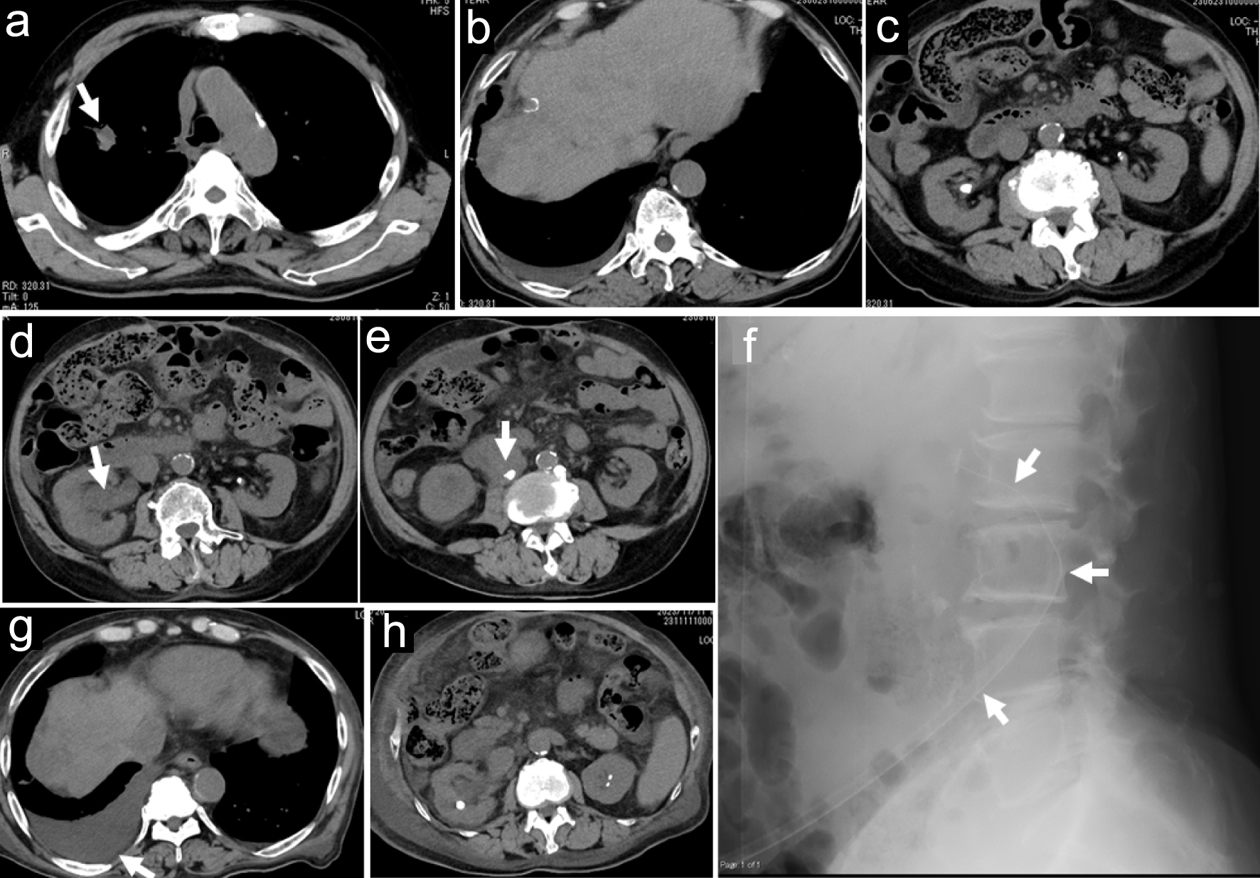
Figure 2. Axial images of computed tomography scans. Nodular lesion in right upper lobe (arrow in a), multiple low-density areas in cirrhotic liver (b), and bilateral nephrolithiasis (c) 3 months before the initial ophthalmic presentation. Ureterolithiasis (arrow in e) with hydronephrosis (arrow in d) on the right side at the time of fever onset, a month before the initial ophthalmic presentation. Lateral view of abdomen X-ray (f) at replacement of a nephrostomy tube (arrows in f) a week after the initial ophthalmic presentation. Stable hepatic cancer in cirrhotic liver (g) and subsiding hydronephrosis (h) 2 months after the initial ophthalmic presentation.

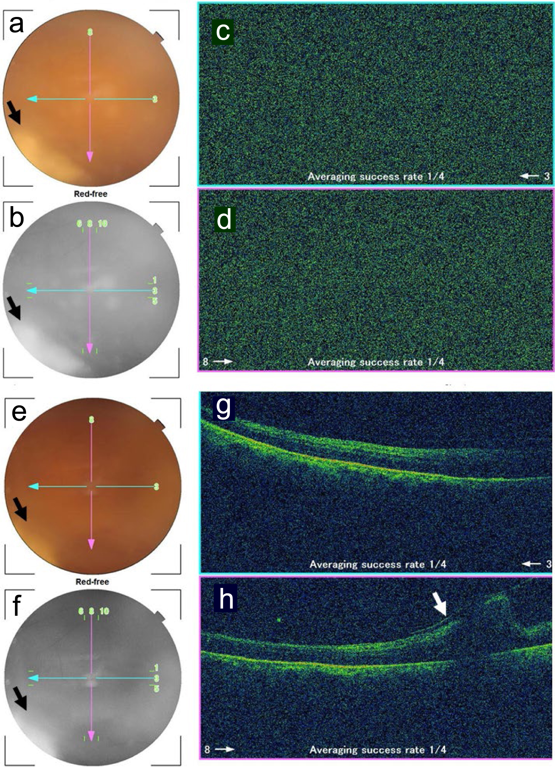
Figure 3. Fundus photographs and optical coherence tomography in the right eye 1.5 months (a-d) and 2 months (e-h) after the initial ophthalmic presentation. a and e: color photographs; b and f: red-free photographs; c and g: horizontal sections designated by blue arrows in fundus photographs; d and h: vertical sections designated by pink arrows in fundus photographs. No images are depicted by optical coherence tomography due to vitreous opacity (c, d) while fluffy retinal infiltrate is visualized in the inferotemporal midperiphery (arrows in a, b, e, f). Note retinal fold (arrow in h) inferior to the macula, caused by vitreoretinal traction.

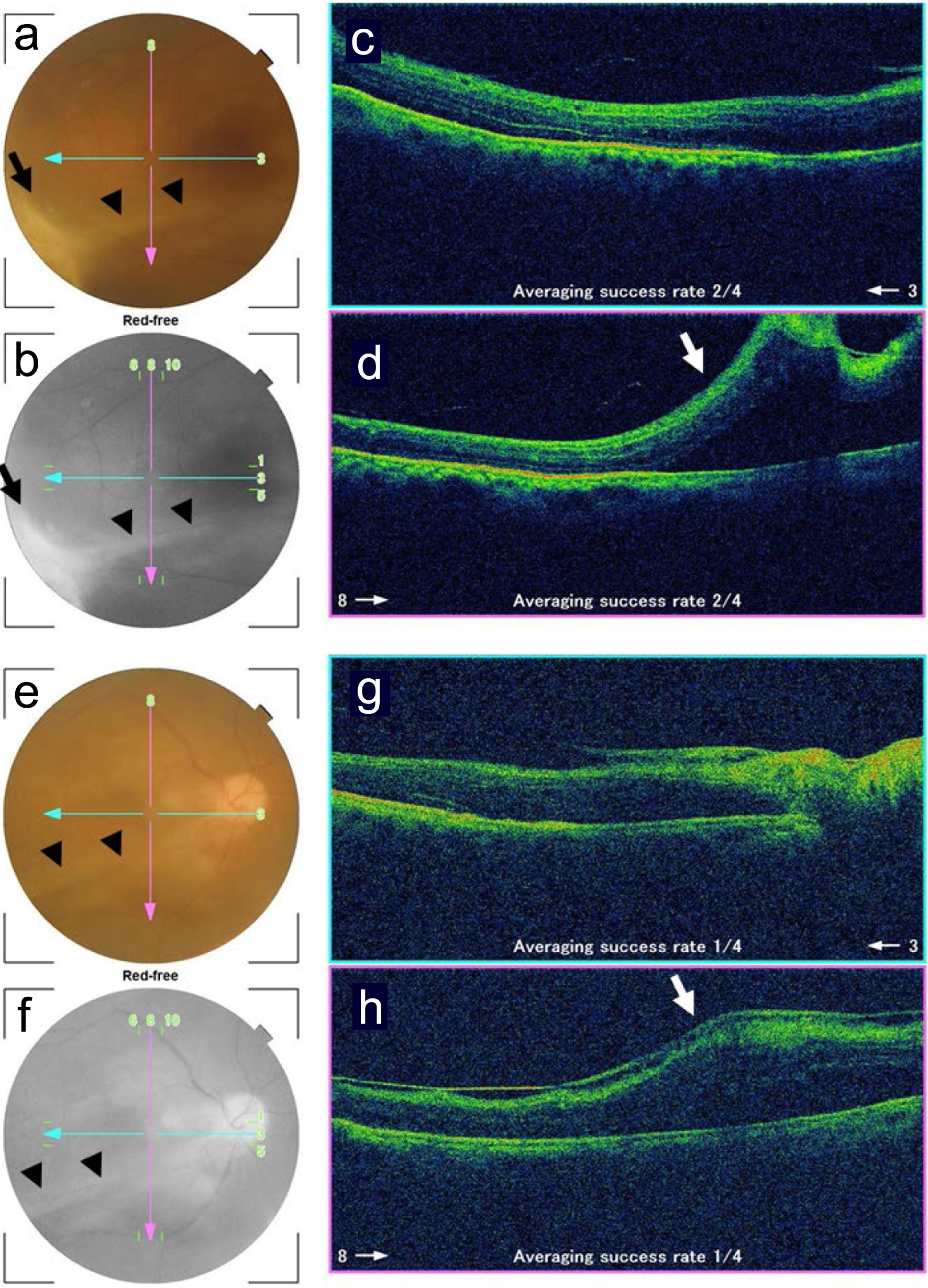
Figure 4. Fundus photographs and optical coherence tomography in the right eye 3.5 months (a-d) and 5 months (e-h) after the initial ophthalmic presentation. a and e: color photographs; b and f: red-free photographs; c and g: horizontal sections designated by blue arrows in fundus photographs; d and h: vertical sections designated by pink arrows in fundus photographs. Note clear vitreous and scarring retinal infiltrate (arrows in a, b) with vitreoretinal adhesion resulting in retinal fold (arrowheads in a, b, e, f, arrows in d, h) inferior to the macula. The retinal fold (arrowheads in e, f) becomes flat in the time course.


