Journal of Medical Cases, ISSN 1923-4155 print, 1923-4163 online, Open Access
Article copyright, the authors; Journal compilation copyright, J Med Cases and Elmer Press Inc
Journal website https://www.journalmc.org

Case Report

Volume 15, Number 11, November 2024, pages 347-353


A Rare Case of Systemic Epstein-Barr Virus-Positive Diffuse Large B-Cell Lymphoma With Hemophagocytic Lymphohistiocytosis in an Immunocompetent Young Man: Potential Diagnostic Pitfall and Therapeutic Challenge

Figures

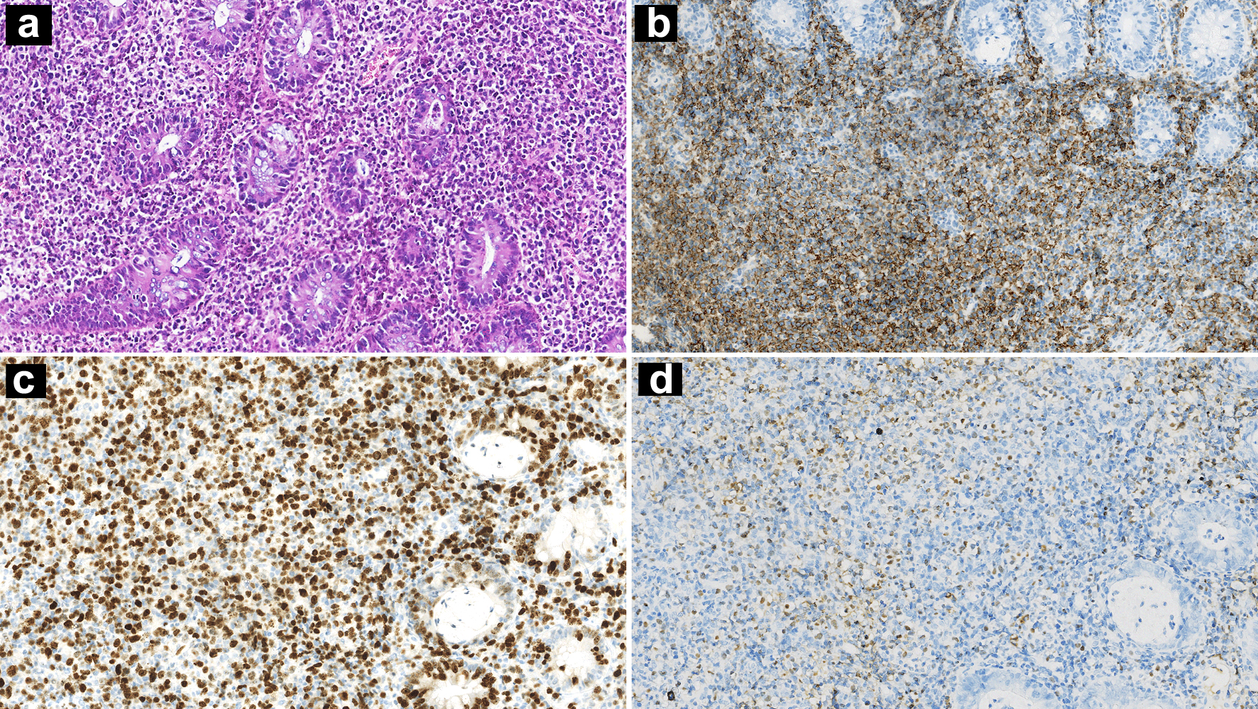
Figure 1.
Figure 1. Nasopharynx biopsy showing diffuse infiltration of atypical lymphoid cells (a: H&E, × 100 magnification). IHC stains showing CD20 positivity (b: × 100 magnification) and a high Ki-67 index (c: × 100 magnification). ISH for EBERs was positive (d: × 100 magnification). H&E: hematoxylin and eosin; IHC: immunohistochemistry; ISH: in situ hybridization; EBER: Epstein-Barr virus-encoded small RNA.
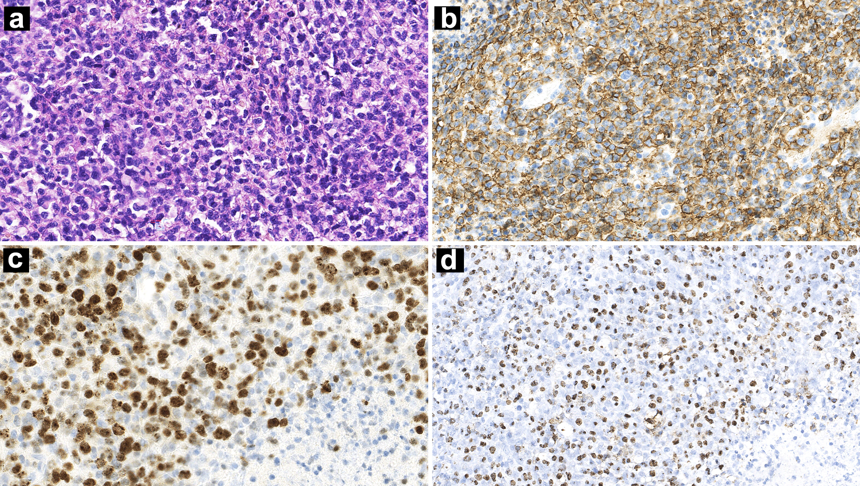
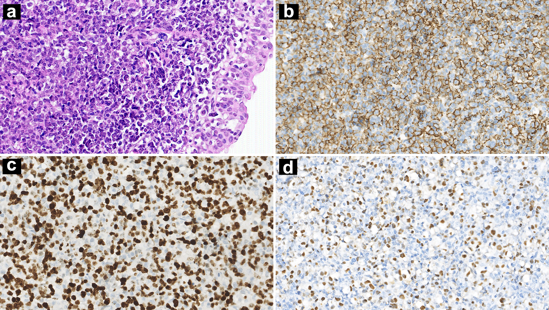
Figure 2.
Figure 2. Lymph nodes biopsy showing diffuse proliferation of lymphoid cells accompanied by necrosis (a: H&E, × 100 magnification). IHC stains showing CD20 positivity (b: × 100 magnification) and a high Ki-67 index (c: × 100 magnification). ISH for EBERs was positive (d: × 100 magnification). H&E: hematoxylin and eosin; IHC: immunohistochemistry; ISH: in situ hybridization; EBER: Epstein-Barr virus-encoded small RNA.
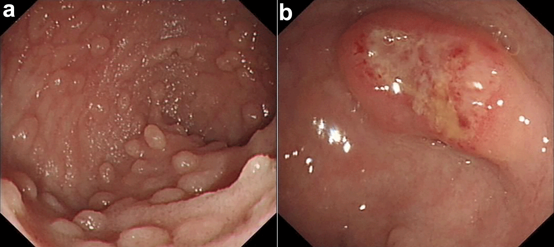
Figure 3.
Figure 3. Endoscopic characteristic of case. Scattered mucosal swellings of varying sizes were found (a). The largest lesion was about 2.5 × 3.0 cm (b).
Figure 4.
Figure 4. Intestinal mucosa biopsy showing diffuse infiltration of large atypical lymphocytes (a: H&E, × 100 magnification). IHC stains showing CD20 positivity (b: × 100 magnification) and high Ki-67 index (c: × 100 magnification). ISH for EBERs was positive (d: × 100 magnification). H&E: hematoxylin and eosin; IHC: immunohistochemistry; ISH: in situ hybridization; EBER: Epstein-Barr virus-encoded small RNA.

Table

Table 1. Differential Diagnosis Among IM, EBV+ LPD with IDD, and EBV+ DLBCL
 
IMEBV+ LPD with IDDEBV+ DLBCL
IM: infectious mononucleosis; EBV+ LPD: EBV-positive lymphoproliferative disorder; IDD: immune deficiency/dysregulation; EBV+ DLBCL: EBV-positive diffuse large B-cell lymphoma; GI: gastrointestinal; CNS: central nervous system; LyG: lymphomatoid granulomatosis; CHL: classic Hodgkin lymphoma.
AgeChildren, young adultsAnyAny
IDD clinical setting-++
Clinical presentationMostly self-limitedRanges from isolated to generalized symptoms> 50% advance stage
Nodal lesion+++
Extranodal lesion+, esp. upper respiratory+, extranodal destructive mass, involve lung, GI tract, CNS, skin, some mimicking LyG+, extranodal destructive mass
Cell proliferationReactive, mixed cell spectrum, foci of immunoblastsPolymorphic infiltrate with full range of muturationMonomorphology, polymorphology, some mimicking CHL or EBV+ LPD
NecrosisSeldomSeldomOften
PhenotypeB-cell derivationB-, T-, and natural killer-cell derivationB-cell derivation
Ki-67 indexVariable, usually lowVariableUsually high
EBV latency32 or 32, few 3
ClonalitySeldom monoclonality< 50% monoclonalityUsually monoclonality