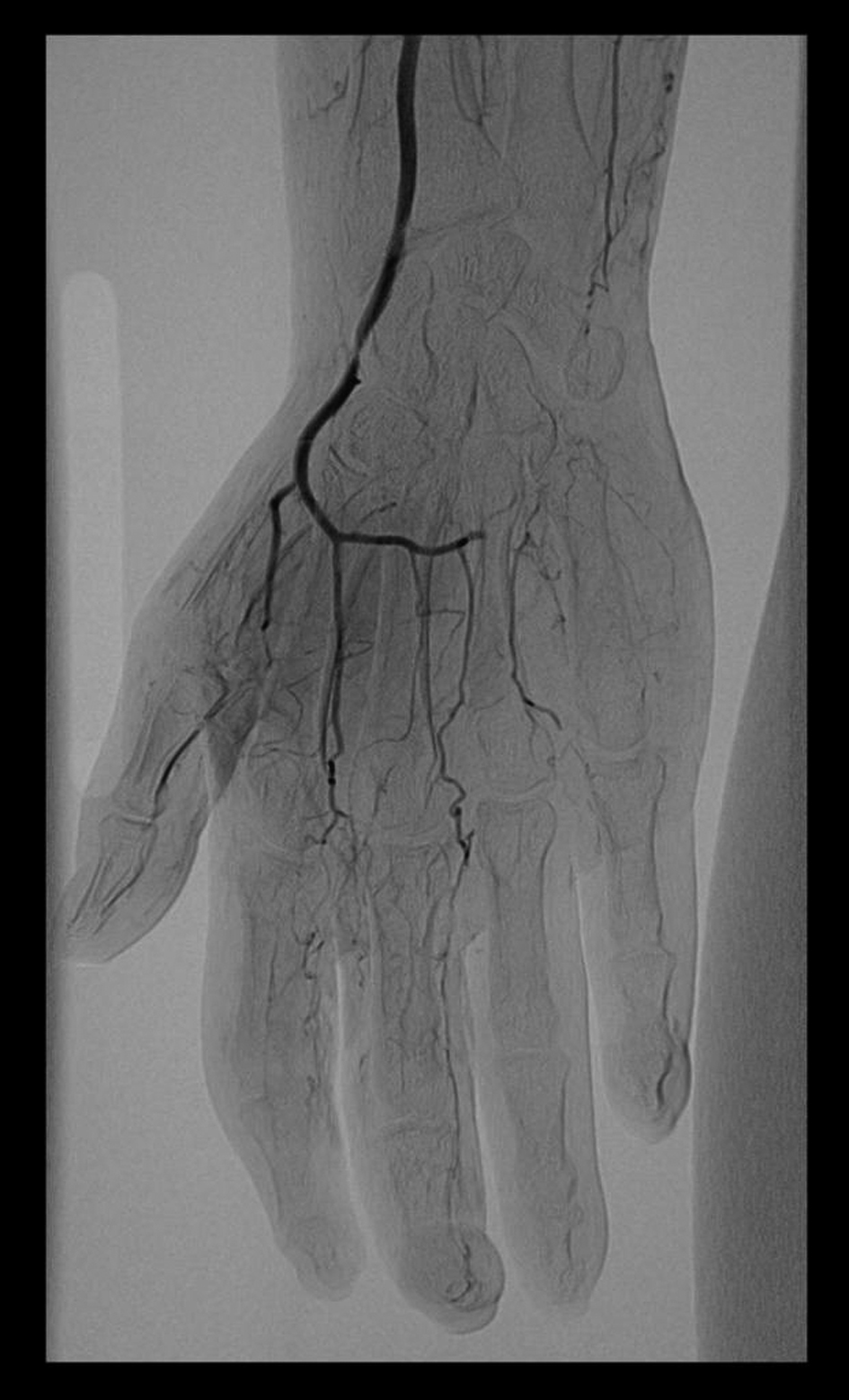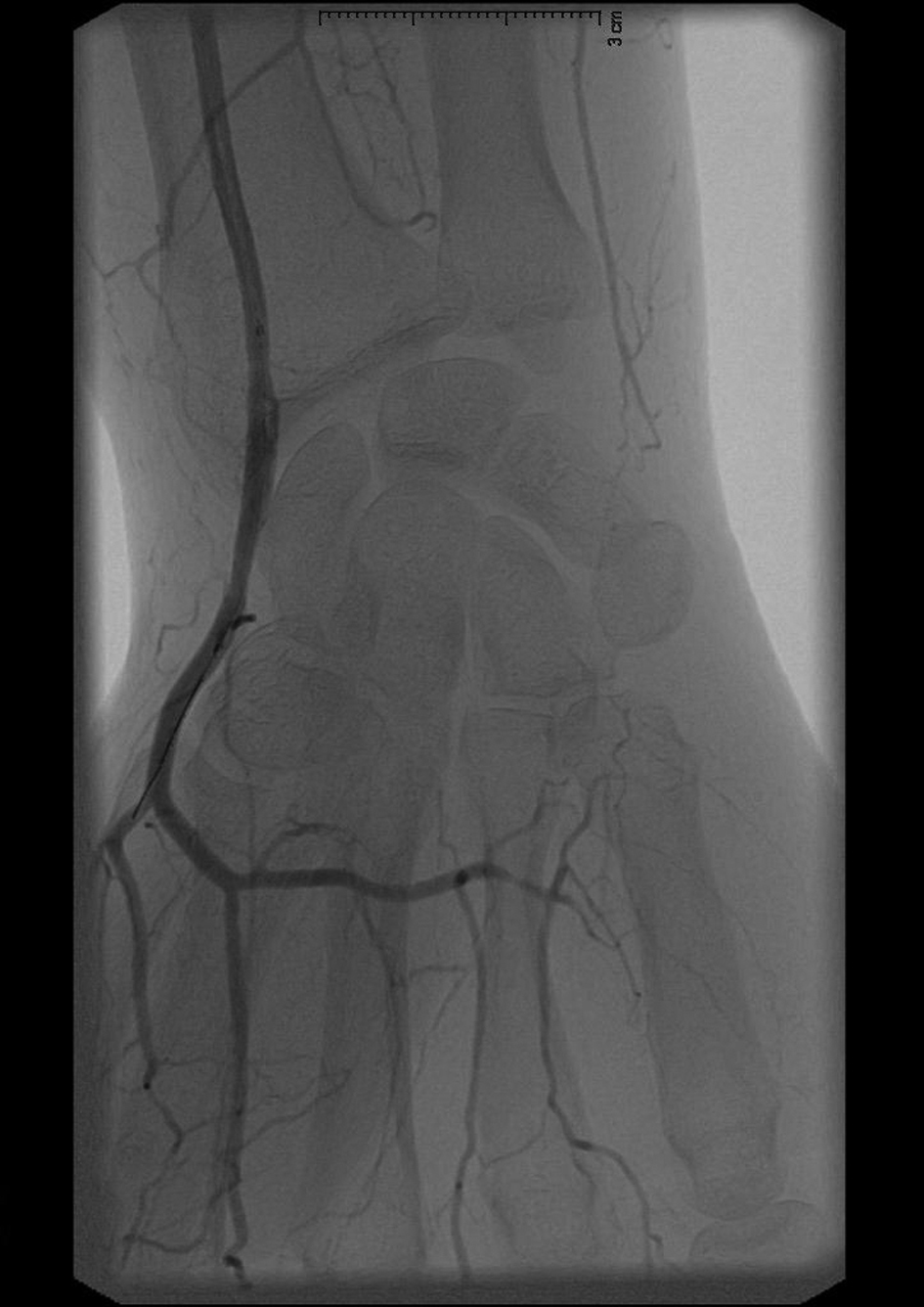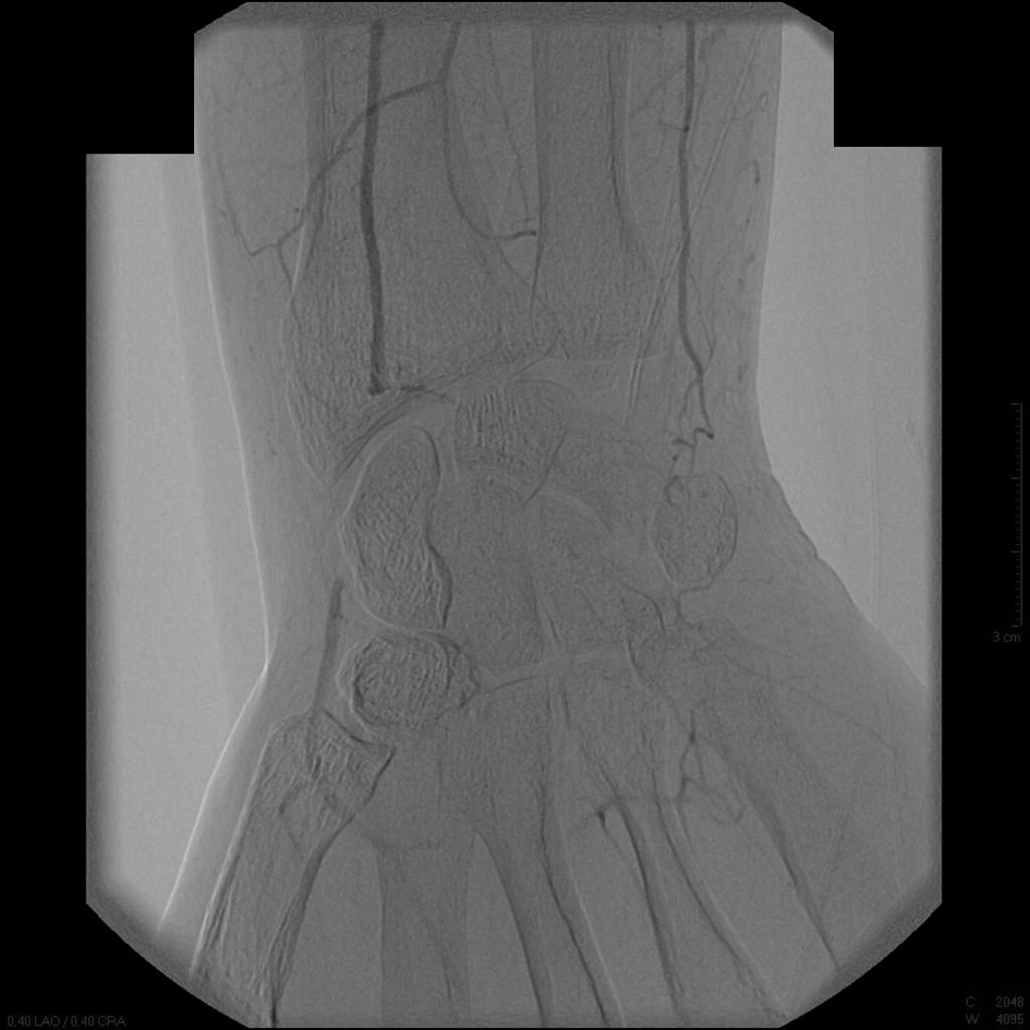
Figure 1. An intra-arterial digital subtraction angiography on the day of second presentation. Occlusion of the right distal radial and ulnar arteries is seen.
| Journal of Medical Cases, ISSN 1923-4155 print, 1923-4163 online, Open Access |
| Article copyright, the authors; Journal compilation copyright, J Med Cases and Elmer Press Inc |
| Journal website http://www.journalmc.org |
Case Report
Volume 4, Number 3, March 2013, pages 185-188
Endovascular Revascularization in Secondary Raynaud’s Phenomenon
Figures