Journal of Medical Cases, ISSN 1923-4155 print, 1923-4163 online, Open Access
Article copyright, the authors; Journal compilation copyright, J Med Cases and Elmer Press Inc
Journal website http://www.journalmc.org

Case Report

Volume 1, Number 3, December 2010, pages 84-86


Intestinal Eosinophilic: Anisakiasis

Figures

Figure 1.
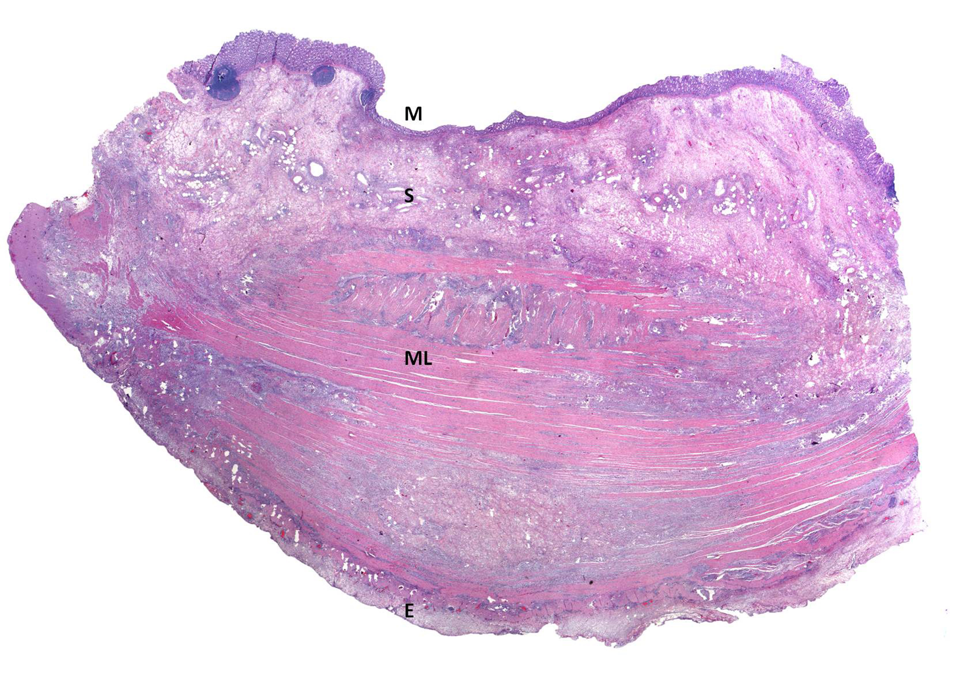
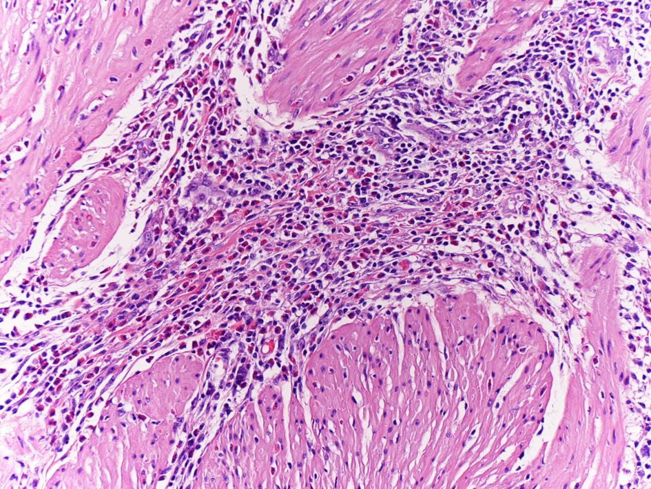
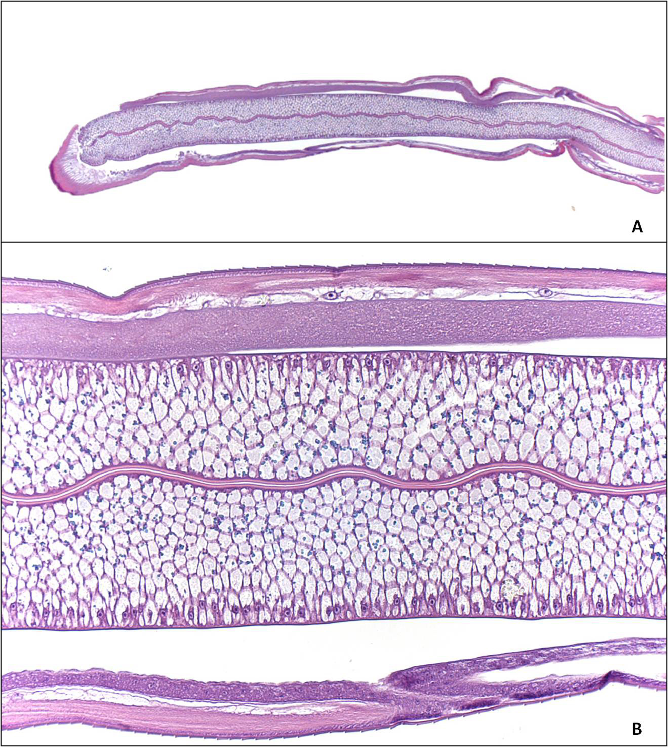
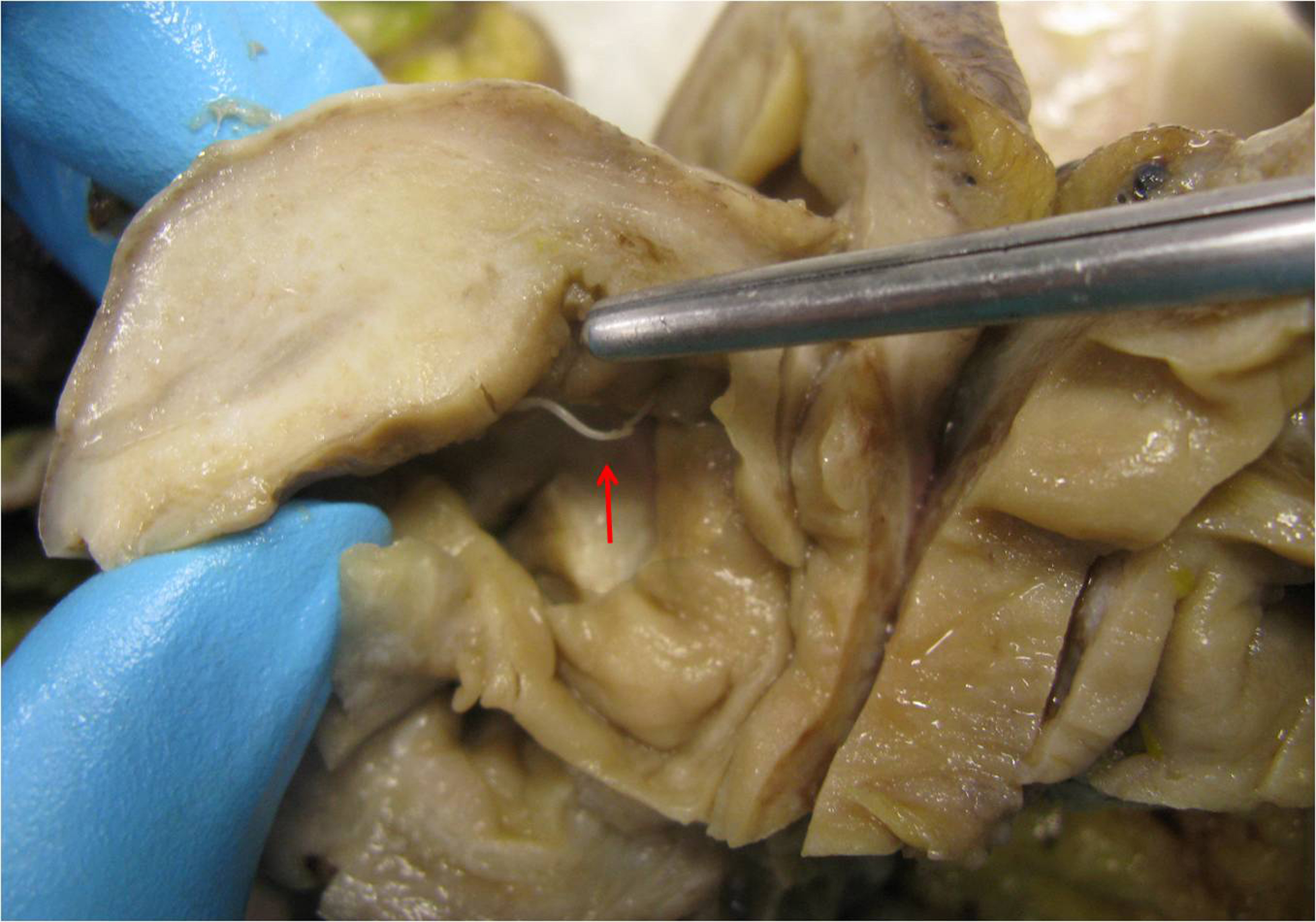
Figure 1. Showed parasite that perforating the bowel mucosa.
Figure 2.
Figure 2. Panoramic histology of the intestinal wall: show mucosa with little erosion (M), edematous submucosa (S), muscle layer with intense inflammatory infiltrate (ML) and the serosa with edema and inflammatory infiltration (E) (hematoxilineand eosine).
Figure 3.
Figure 3. Presenting an intense inflammatory infiltrate with eosinophils (magnification x 40,Hematoxiline and Eosine).
Figure 4.
Figure 4. Histological examination of the parasitic nematode, showing a thick cuticle, neural cord, and gastrointestinal structure (A, B) (hematoxiline andeosine).