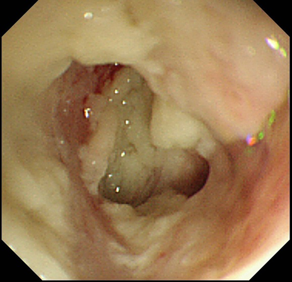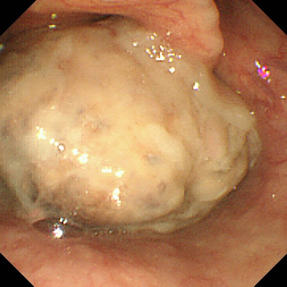
Figure 1. The upper gastrointestinal endoscopy with thin scope showed a white-colored, movable mass lesion occupying the lower pharynx.
| Journal of Medical Cases, ISSN 1923-4155 print, 1923-4163 online, Open Access |
| Article copyright, the authors; Journal compilation copyright, J Med Cases and Elmer Press Inc |
| Journal website http://www.journalmc.org |
Case Report
Volume 3, Number 3, June 2012, pages 211-213
Expanding Growth and Pharyngeal Mass Formation in Elderly Patient With Esophageal Cancer: Unusual Manifestation
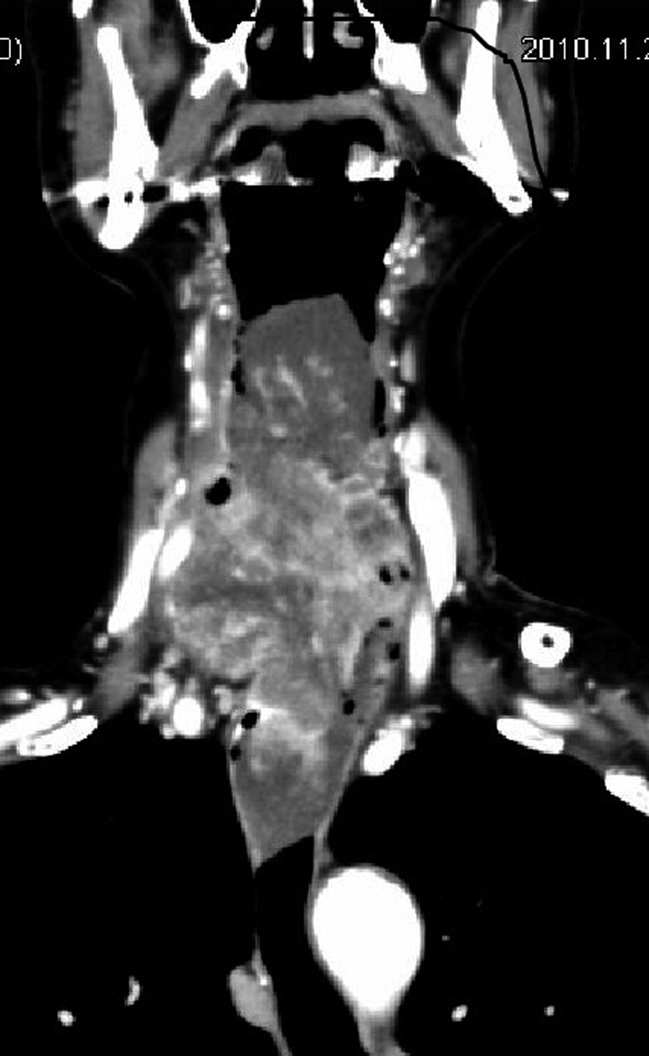
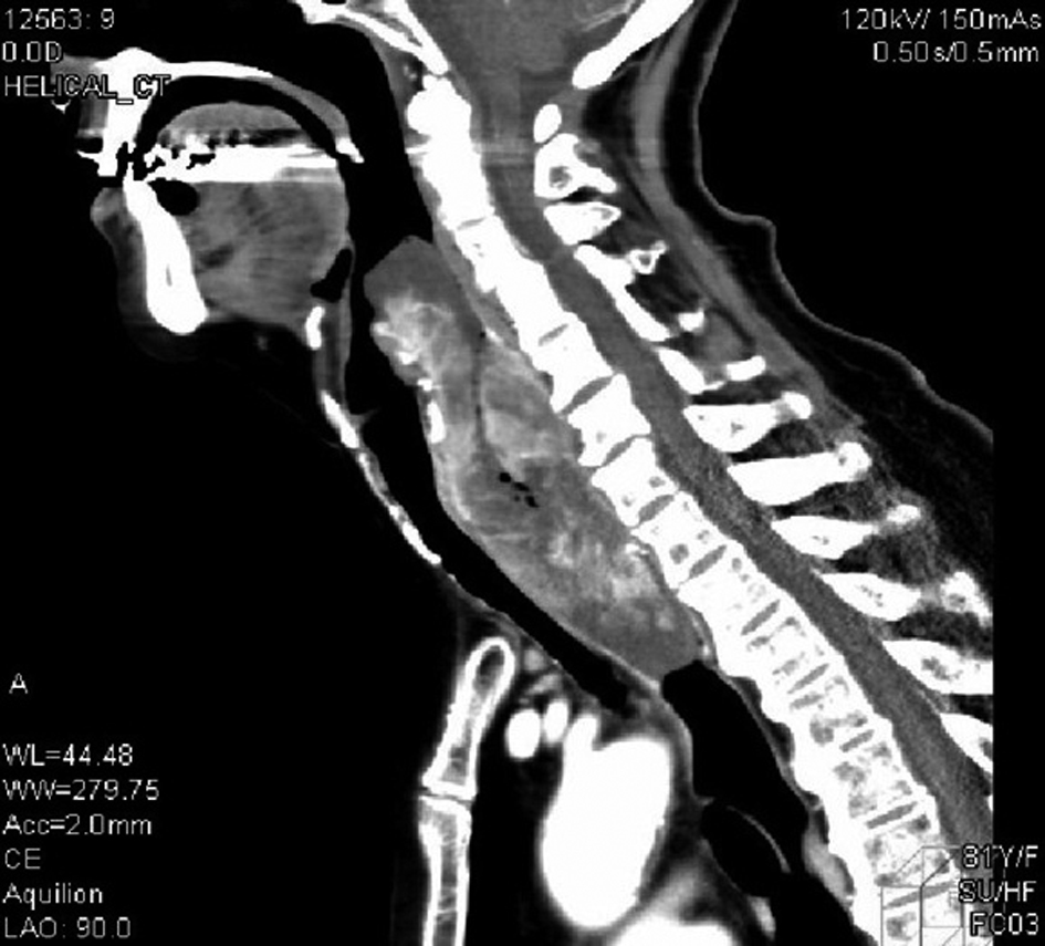
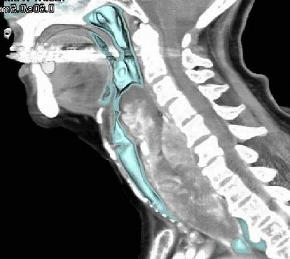
Figures