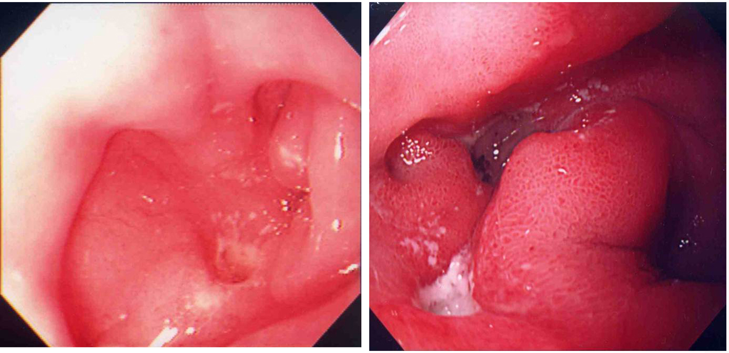
Figure 1. Past endoscopic findings; 19 years old (left) and 32 years old (right).
| Journal of Medical Cases, ISSN 1923-4155 print, 1923-4163 online, Open Access |
| Article copyright, the authors; Journal compilation copyright, J Med Cases and Elmer Press Inc |
| Journal website http://www.journalmc.org |
Case Report
Volume 3, Number 4, August 2012, pages 243-246
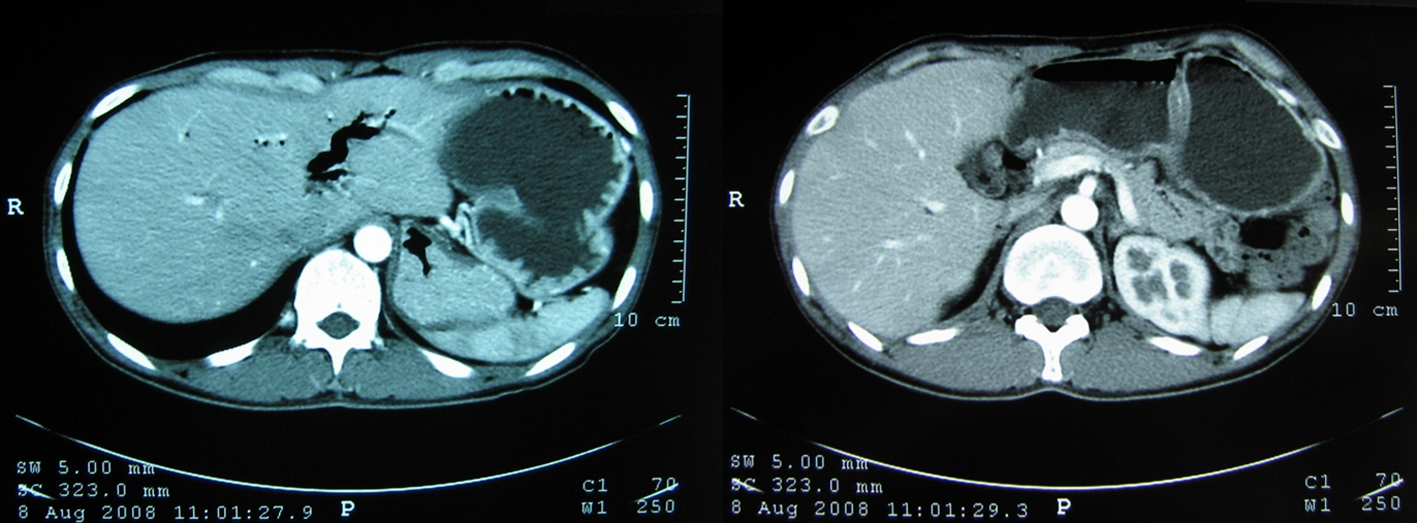
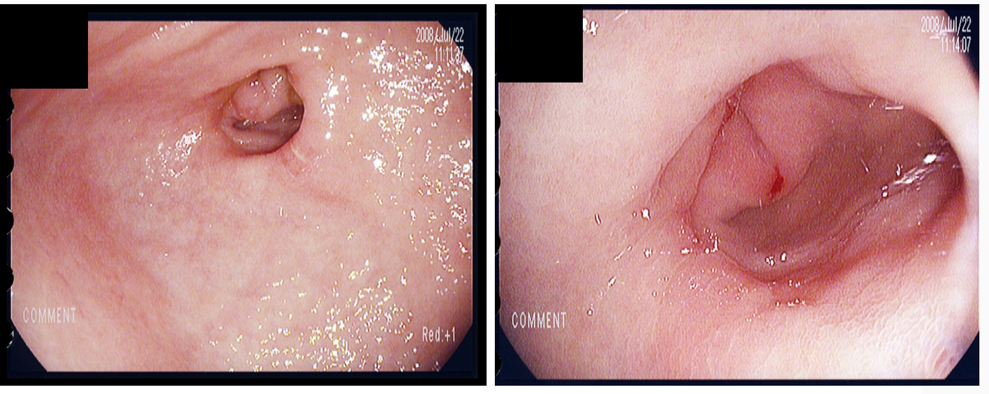
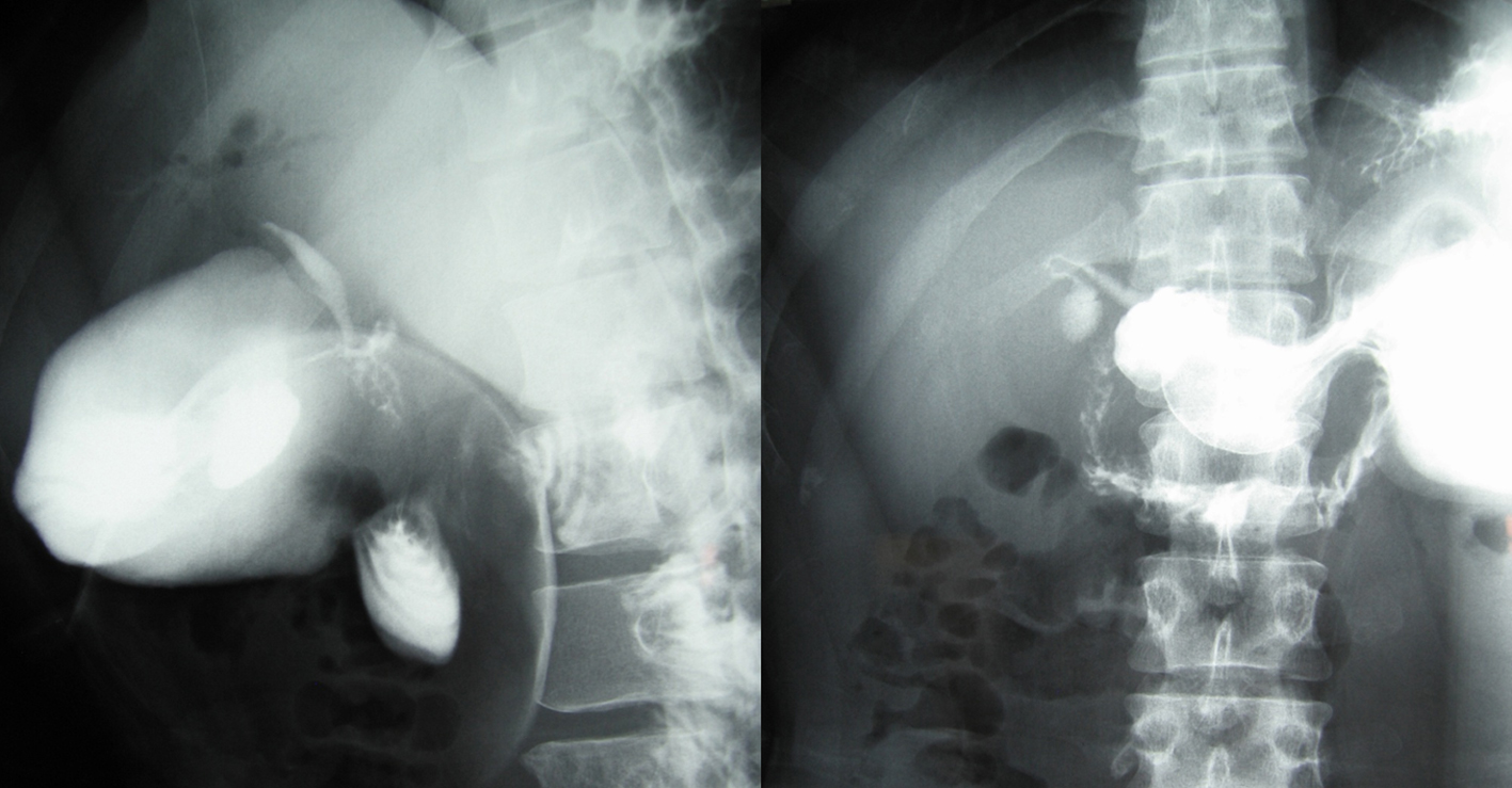
Choledochoduodenal Fistula Associated With Recurrent Peptic Ulcer
Figures
Table
| Cell count | |
|---|---|
| RBC | 511 × 104/µl |
| Hb | 15.9 g/dl |
| Ht | 46.5% |
| Plt | 26.6 ×10⁴/µl |
| WBC | 7,800 /µl |
| Blood gas analysis (room air) | |
| pH | 7.56 |
| paCO2 | 74 mmHg |
| paO2 | 70 mmHg |
| HCO3 | 66.3 mmol/l |
| SaO2 | 94.6% |
| Chemistry | |
| TP | 8.1 g/dl |
| Alb | 5.4 g/dl |
| AST | 20 IU/l |
| ALT | 16 IU/l |
| LDH | 149 IU/l |
| AIP | 248 IU/l |
| γ-GTP | 23 IU/l |
| ChE | 5.44 U/ml |
| BUN | 61.8 mg/dl |
| Cr | 3.44 mg/dl |
| UA | 13.5 mg/dl |
| Na | 145 mEq/l |
| K | 2.9 mEq/l |
| Ca | 4.7 mEq/l |
| FBS | 96 mg/dl |
| CRP | 0.76 mg/dl |