Journal of Medical Cases, ISSN 1923-4155 print, 1923-4163 online, Open Access
Article copyright, the authors; Journal compilation copyright, J Med Cases and Elmer Press Inc
Journal website http://www.journalmc.org

Case Report

Volume 3, Number 4, August 2012, pages 250-253


Intravitreal Ranibizumab for the Treatment of Choroidal Neovascularization in Best’s Vitelliform Macular Dystrophy

Figures

Figure 1.
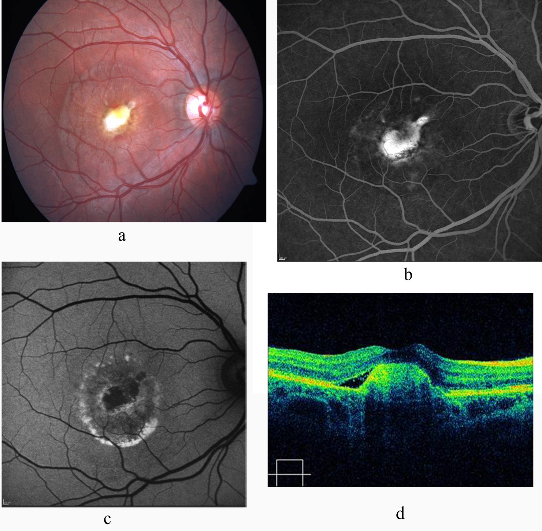
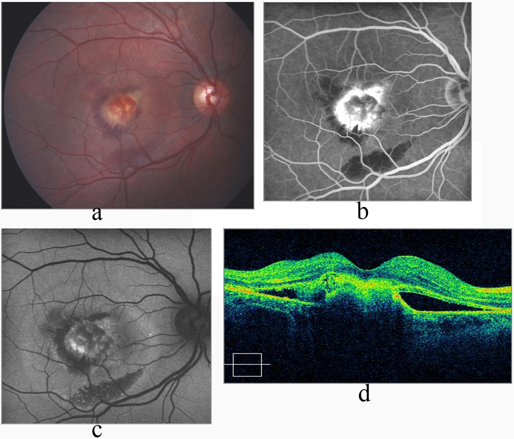
Figure 1. a: Macular edema and hemorrhage in the right eye in Best’s disease; b: FA shows increased hyperfluoresence due to CNV, and hypofluorescence due to hemorrhage in the late phase; c: FAF image shows hypoautofluorescence due to subretinal classical CNV, and hemorrhage; d: OCT shows hyperreflective submacular CNV with subretinal fluid 6 months after the last injection of intravitreal ranibizumsb.
Figure 2.
Figure 2. a: Color fundus photograph shows scarred CNV with resolution of the subretinal fluid and hemorrhage; b: FA shows absence of leakage and staining of the CNV; c: FAF image shows hypoautofluorescent scar with hyperautofluorescent deposits arround it; d: Significant reduction of the subretinal fluid and retinal thickness on OCT.