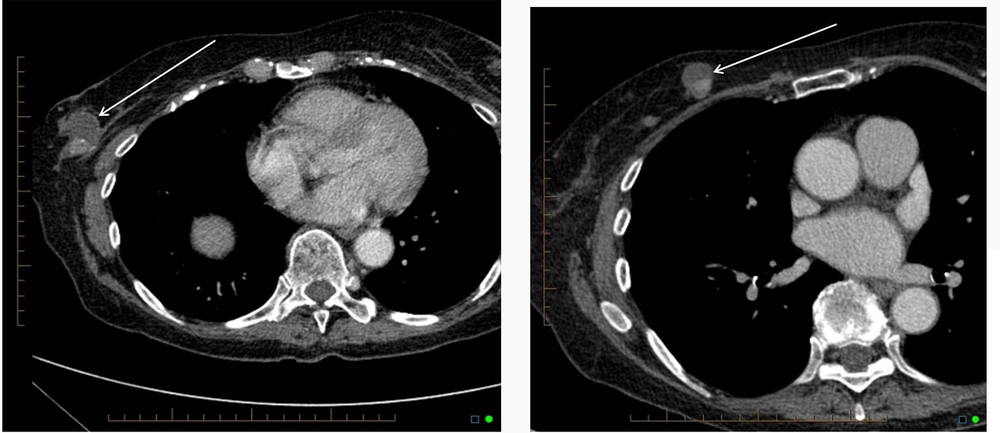
Figure 1. CT scan of the chest showing two partially cystic, partially solid mass lesions in the right breast.
| Journal of Medical Cases, ISSN 1923-4155 print, 1923-4163 online, Open Access |
| Article copyright, the authors; Journal compilation copyright, J Med Cases and Elmer Press Inc |
| Journal website http://www.journalmc.org |
Case Report
Volume 3, Number 4, August 2012, pages 270-273
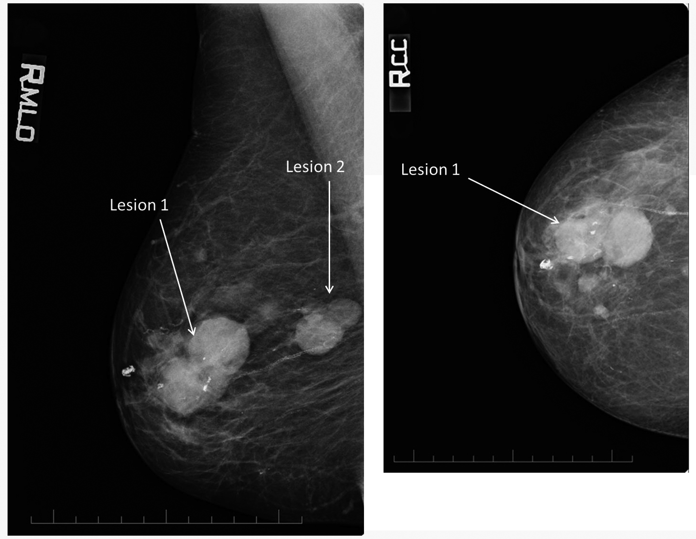
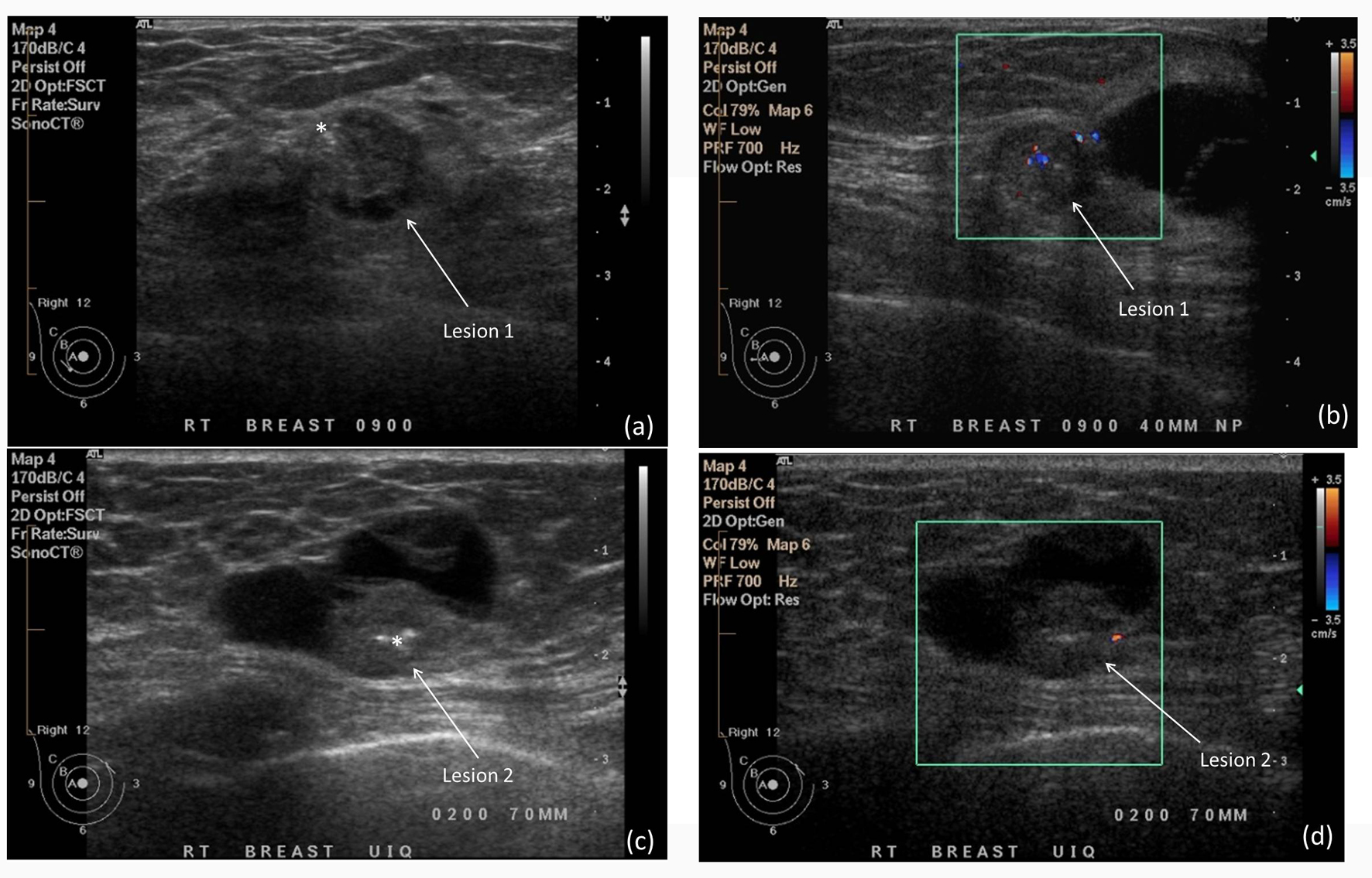
Unusual Mammographic and Ultrasound Findings in a Patient With Ductal Carcinoma in Situ (DCIS)
Figures