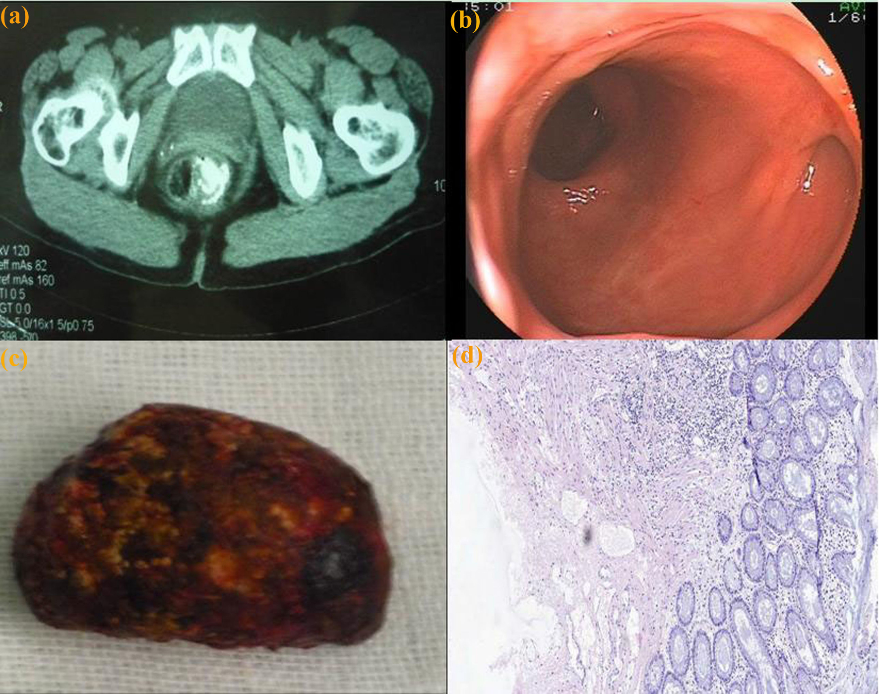
Figure 1. (a) Computer tomography: a 34 x 23 mm lesion, in the right supralevator space, was identified. The lesion was communicated with rectal lumen at the level of the staple line. (b) Colonoscopy view: an isolated diverticulum cavity in the rectum. (c) Surgical specimen: an impacted fecalith in the rectal diverticulum cavity. (d) Histologic examination: the entire rectal layer with the mucosa, submucosa, and proper muscle (H and E stain, x 5).