Journal of Medical Cases, ISSN 1923-4155 print, 1923-4163 online, Open Access
Article copyright, the authors; Journal compilation copyright, J Med Cases and Elmer Press Inc
Journal website http://www.journalmc.org

Case Report

Volume 3, Number 6, December 2012, pages 375-378


Unusual Presentation of a Dysembryoplastic Neuroepithelial Tumor Mimicking a Pontomesencephalic Cavernous Hemangioma

Figures

Figure 1.
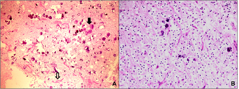
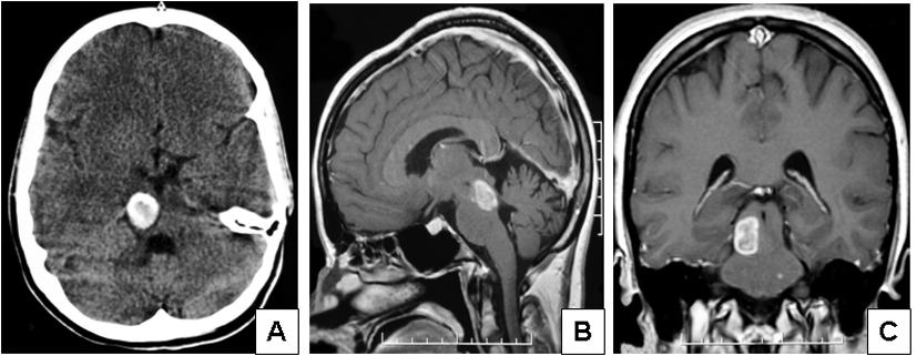
Figure 1. Imaging features of the case of dysembryoplastic neuroepithelial tumor presenting in the pontomesencephalic area. (A) Contrast enhanced CT scan showed a large, well-demarcated, subcortical hyperdense lesion in the right mesencephalon with extension into the tegmentum pontis. (B, lateral) and (C, coronal) MRI examination revealed a pontomesencephalic pseudocystic lesion that was hyperintense on T1 weighted images and showed less enhancement on T2 weighted images extending within the brain stem and reaching the red nucleus, the superior colliculi and the superior cerebellar tract.
Figure 2.
Figure 2. Microscopic features of the case of dysembryoplastic neuroepithelial tumour presenting in the pontomesencephalic area. (A) Low power view demonstrates a loosely structured mucinous background with numerous psammomatous calcifications (solid arrow) and areas of hamartomatous vasculature (open arrow) (H&E, x 100). (B) Medium power view reveals randomly distributed mature-appearing oligodendroglia-like cells suspended in a partially microcystic mucinous background (“floating-neurons”) (H&E, x 200).