Journal of Medical Cases, ISSN 1923-4155 print, 1923-4163 online, Open Access
Article copyright, the authors; Journal compilation copyright, J Med Cases and Elmer Press Inc
Journal website http://www.journalmc.org

Case Report

Volume 3, Number 5, October 2012, pages 326-328


Think Heterotopic: A Case Report of Heterotopic Pregnancy Detected on Thorough Ultrasonography

Figures

Figure 1.
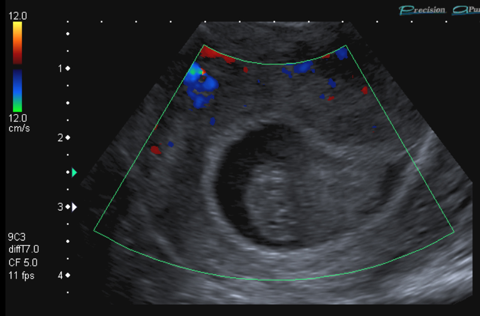
Figure 1. Transvaginal ultrasound scan showing non-viable intrauterine gestation with probable fetal pole without yolk sac nor fetal cardiac pulsation. Crown lump length was estimated at 11 mm (7+ weeks gestation). Visualisation of content of gestational sac is hard due to haemorrhagic material.
Figure 2.
Figure 2. Transvaginal ultrasound scan showing a large irregular heterogenous mass of 48 mm x 31 mm x 21 mm with 12 mm x 6 mm x 5 mm probable yolk sac consistent with right ectopic pregnancy.
Figure 3.
Figure 3. Transvaginal ultrasound scan showing free fluid in the pouch of Douglas indicated by increased echogenicity.