Journal of Medical Cases, ISSN 1923-4155 print, 1923-4163 online, Open Access
Article copyright, the authors; Journal compilation copyright, J Med Cases and Elmer Press Inc
Journal website http://www.journalmc.org

Case Report

Volume 4, Number 3, March 2013, pages 142-145


Laryngeal Sarcoidosis Causing Acute Airway Obstruction: A Rare Sequela of a Rare Entity

Figures

Figure 1.
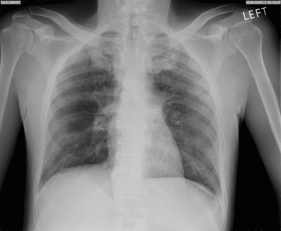
Figure 1. Chest X-ray showing non-specific interstitial edema.
Figure 2.
Figure 2. Computed Tomography Scan of the neck (Axial view) showing submucosal infiltration of the supraglottic larynx and left vocal cord fixation.
Figure 3.
Figure 3. Computed Tomography Scan of the neck (Sagittal reconstruction) showing acute airway narrowing.
Figure 4.
Figure 4. Histopathology of the supraglottis mass (low power) showing squamous epithelium (black arrow) and underlying soft tissue showing multiple non-caseating granuloma (white arrow).
Figure 5.
Figure 5. Histopathology of the supraglottis mass (high power) showing Langhans cells.