Journal of Medical Cases, ISSN 1923-4155 print, 1923-4163 online, Open Access
Article copyright, the authors; Journal compilation copyright, J Med Cases and Elmer Press Inc
Journal website http://www.journalmc.org

Case Report

Volume 4, Number 1, January 2013, pages 4-8


Severe Pneumonia Caused by Streptococcus Pneumoniae and Haemophilus Influenzae Coinfection in an Elderly Patient: A Case Report

Figures

Figure 1.
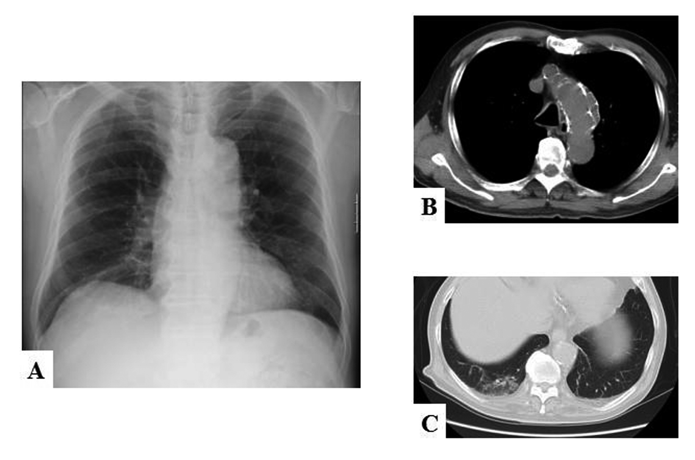
Figure 1. (A) Chest X-ray film and (B, C) computed tomograms of the patient on admission. (A) Mild consolidation is apparent along the right lower lobe branch of the pulmonary artery. The silhouette sign is negative, suggesting the right lower lobe is the site of the pneumonia. (B) Computed tomogram shows an old dissecting aneurysm of the aortic arch (white arrows). (C) The pneumonia mainly affects S10 of the right lower lobe.
Figure 2.
Figure 2. Chest X-ray films on (A) day 5 and (B) day 10. (A) Bilateral infiltration of the lung, which was not present on day 3, is now apparent. (B) The radiographic findings have improved considerably by 4 days after intubation (day 10). Although some infiltration remains, the SpO2 is now 100% on 30% FiO2, accordingly we extubated him.