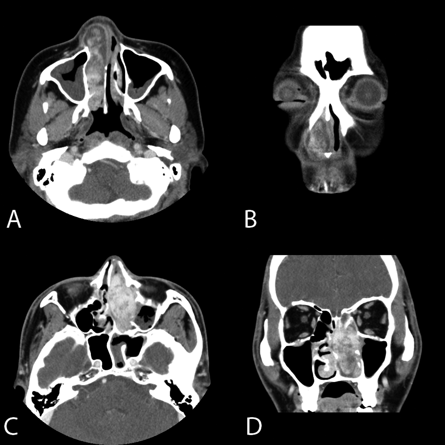
Figure 1. Axial (A) and coronal (B) enhanced computed tomography of the paranasal sinuses (PNS CT) of case 1 demonstrate a 3 × 5 cm sized lobuated soft tissue opacity occupying the entire right nasal cavity with intense enhancement. The lesion was well circumscribed without any evidence of bony destruction. Axial (C) and coronal (D) PNS CT of case 2 show a soft tissue opacity with intense enhancement occupying the entire left nasal cavity. The nasal septum was deviated to the right side. However, there is no evidence of bony destruction.

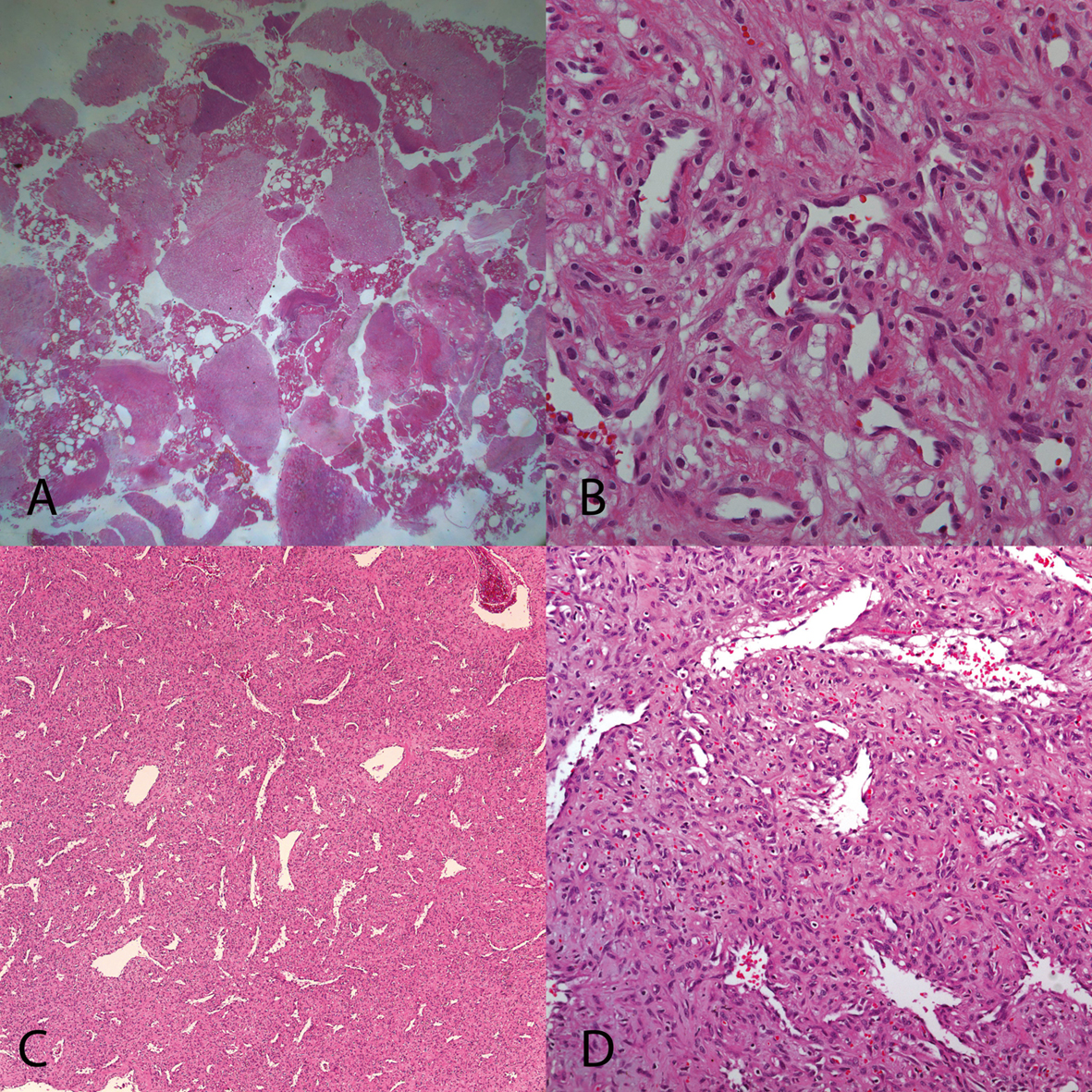
Figure 2. In histopathological examination of case 1 (A, B) and case 2 (C, D), multiple polypoid fragments of angiomatous tissue protruding above the surrounding mucosa were seen. The mass was covered with pseudostratified ciliated epithelium showing focal squamous metaplasia and ulceration below which can be seen profound inflammation in the connective tissue. The submucosal lamina propria contained lobular proliferated capillaries with lumina of varying sizes surrounded by an edematous fibromixoid stroma. There was no evidence of mitotic activity or malignancy. These histopathological findings were consistent with a lobular capillary hemangioma LCH. (A, C: H & E, × 40, B, D: H & E, × 200).

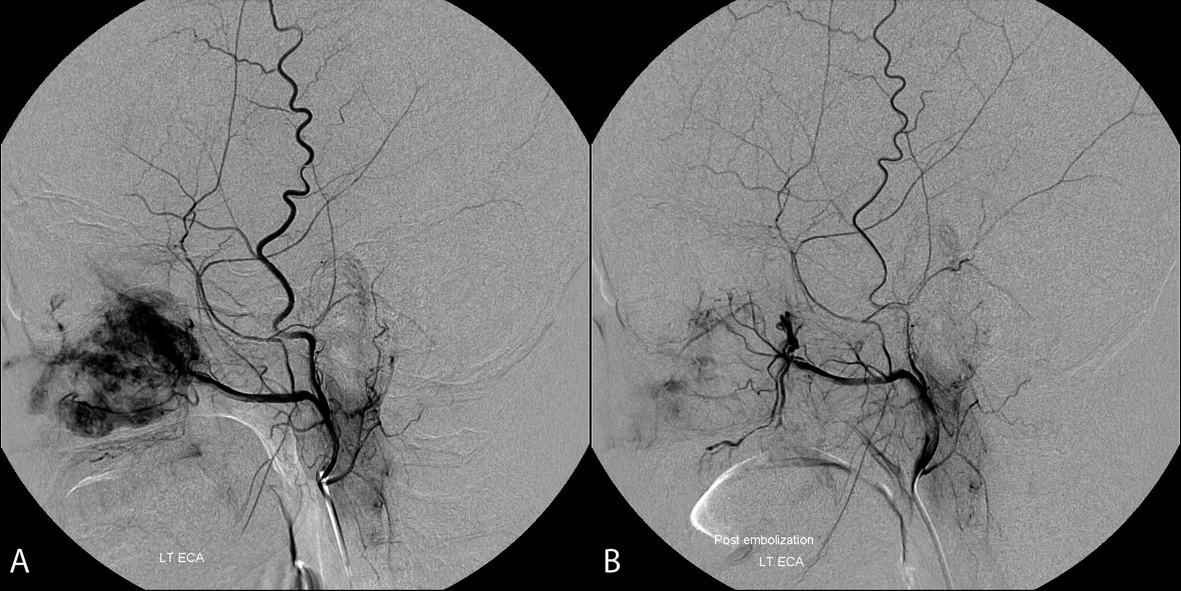
Figure 3. Carotid angiogram of nasal mass in case 2 shows mass (arrow) lesion staining in the left nasal cavity, supplied by internal maxillary artery of left external carotid artery (A). After successful embolization by polyvinyl alcohol, no more staining lesion was observed (B).


