Journal of Medical Cases, ISSN 1923-4155 print, 1923-4163 online, Open Access
Article copyright, the authors; Journal compilation copyright, J Med Cases and Elmer Press Inc
Journal website http://www.journalmc.org

Case Report

Volume 2, Number 1, February 2011, pages 7-12


A Case of Mucosa-Associated Lymphoid Tissue Lymphoma (MALToma) That Developed From Lungs and Stomach

Figures

Figure 1.
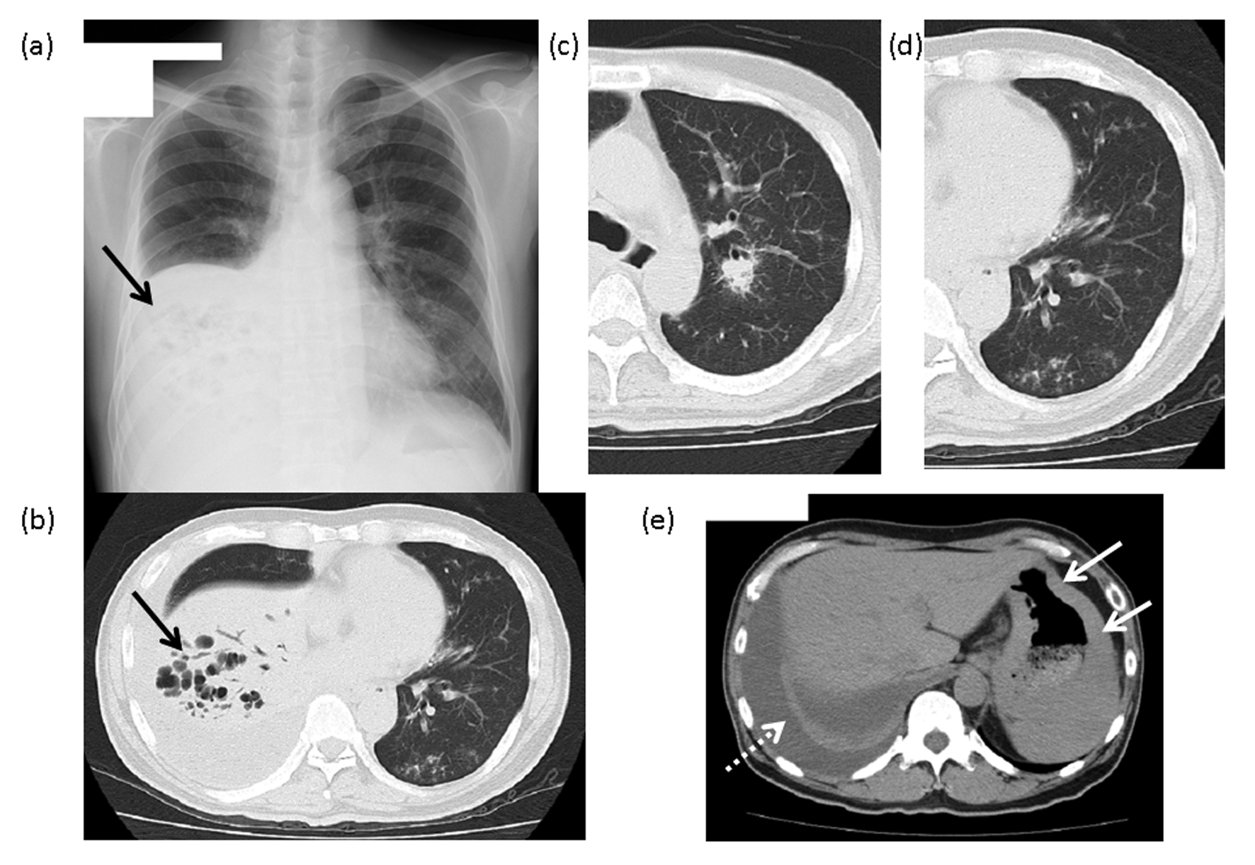
Figure 1. The findings of chest X-ray and computed tomography (CT). (a) Chest X-ray: An area of reduced permeability, with multiple cysts, is visible in the lower right lobe (arrow). (b) Chest CT: An infiltrative shadow with manifest bronchiectasis can be seen in the right lower lobe (arrow). (c) A nodular lesion with an air bronchogram is visible around the center side of the bronchus of the right upper lobe. (d) Multiple ground-glass opacities and granular lesions are visible around the bronchus on the periphery of the left lower lobe. (e) A right pleural effusion (dashed line arrow) and the gastric wall with manifest thickening (arrow) can be seen on this image.
Figure 2.
Figure 2. The cytology of the pleural effusion. Large numbers of cells were detected, which were class IV by Papanicolaou staining, larger than lymphocytes, basophilic by Giemsa staining and with cellular nuclei that were enlarged and existed deviating from center. This led us to suspect multiple myeloma, leukemia or malignant lymphoma.
Figure 3.
Figure 3. The brush cytology of the bronchus in the right lower lobe. Cells were detected which were class V by Papanicolaou stain, and similar to those in the pleural effusion.
Figure 4.
Figure 4. Trans-bronchial lung biopsy of the right lower lobe. A massive invasion of lymphocytes and a lymphoepithelial lesion (LEL) is presented, by which MALToma was suspected.
Figure 5.
Figure 5. Chromosome analysis of the patient’s pleural effusion. Chromosome banding in the pleural effusion, performed in Nippon Medical School Hospital, identified the aberration of t(11;18)(q21;q21.3).
Figure 6.
Figure 6. Gastric biopsy in Nippon Medical School Hospital. (a) HE staining: as well as a specimen of TBLB, there was a manifest lymphoid invasion and a lymphoepithelial lesion (LEL). (b) CD20 staining: manifest invasion of CD20-positive B lymphocytes. (c) Keratin staining: destruction of duct configuration by lymphocytes.

Table

Table 1. Results of Blood Examination, Urinalysis, Urinary Sediment and PE Findings
 
(CBC), (complete blood count); WBC, white blood cell; RBC, red blood cell; Hb, hemoglobin; Ht, hematocrit; Plt, blood platelet; (HG), hemogram; stab, stab cell; seg, segmented cell; lympho, lymphocyte; mono, monocyte; eosino, eosinophil; baso, basophil; BBC, blood biochemistry; AST, aspartate aminotransferase; ALT, alanine aminotransferase; LDH, lactate dehydrogenase; BUN, blood urea nitrogen; Cre, creatinine; UA, uric acid; TP, total protein; T-cho, total cholesterol; TG, triglyceride; Glu, glucose; CRP, C-reactive protein; CEA, carcinoembryonic antigen; SCC, squamous cell carcinoma related antigen; NSE, neuron specific enolase; IgG, immunogloblin G; IgM , immunogloblin M; IgE, immunoglobulin E; IEP, Immuno-electrophoresis; sLI-2R, serum interleukin II receptor; (ABG), (arterial blood gas analysis); BE, base excess; SaO2, Oxygen saturation; PE, pleural effusion; BJP, Bence-Jones protein; ADA, adenosine deaminase.
(CBC)(BBC)(Urinalysis)
WBC19500/mmGOT14 U/lIgA115 mg/dlPH6
RBC448×104/mmGPT10 U/lIgG2022 mg/dlProtein-
Hb14.2 g/dlLDH243 U/lIgM570 mg/dlSugar-
Ht41.7%TP8.4 g/dlIgE13Occult blood±
MCV93T-cho115 mg/dlIEPIgM-κ TypeBJP-
MCH31.7TG69 mg/dlIgG-κ Type-sediment-
MCHC34.1%HDL-cho30 mg/dlsIL-2R1740 U/dlRBC1-4/HPF
Plt41.7×104/mmNa133 mEqWBC< 1/HPF
(Hemogram)K4.6 mEq(ABG)
Stab3%Cl102 mEqPH7.454(PE)
Seg86%Glu110 mg/dlPCO236.2 mmHgPH7.2
Lympho4.5%CRP4.73 mg/dlPO273 mgHgprotein6.0g/dl
Mono4%CEA1.7 ng/dlHCO3-24.8 mmol/lADA163IU/L
Eosino1.5%SCC0.6 ng/dlBE1.3 mmol/lCellneutrophil 12%
Baso1%NSE8.6 ng/dlSaO295.4%Fractionationlymphocyte 88%