Journal of Medical Cases, ISSN 1923-4155 print, 1923-4163 online, Open Access
Article copyright, the authors; Journal compilation copyright, J Med Cases and Elmer Press Inc
Journal website http://www.journalmc.org

Case Report

Volume 4, Number 1, January 2013, pages 43-45


A Rare Presentation of Stroke: Basilary Artery Dissection

Figures

Figure 1.
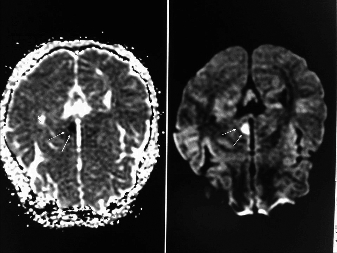
Figure 1. On evaluation of cranial diffusion-weighted magnetic resonance image (MRI), a lesion region slightly hypodense in T1A and slightly hyperdense in T2A series and primarily thought to be acute ischemic was observed in the left thalamus.
Figure 2.
Figure 2. A three-dimensional computed tomographic angiography (CTA) of the head showed narrowing of the basilar artery from the distal ends of the anterior inferior cerebellar artery (AICA) branches to posterior cerebellar artery (PCA) in volume rendered (VR) images. There was also narrowing in the left PCA P1 segment as well as right PCA P1 and P2 segments.
Figure 3.
Figure 3. Vertebrobasilar Digital Substraction Angiography (DSA) and reduction in artery calibration at AICA exit in the distal end of the basilary artery was seen following the vertebtal artey injection in anterior posterior plane.
Figure 4.
Figure 4. Thinning of the lumen (String sign), flap and filling of the false lumen observed along with the real vessel calibration in lateral projection.