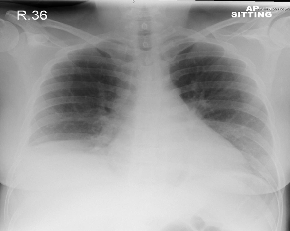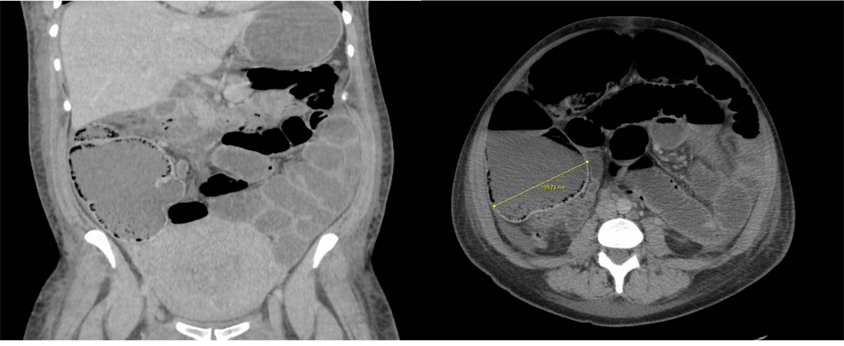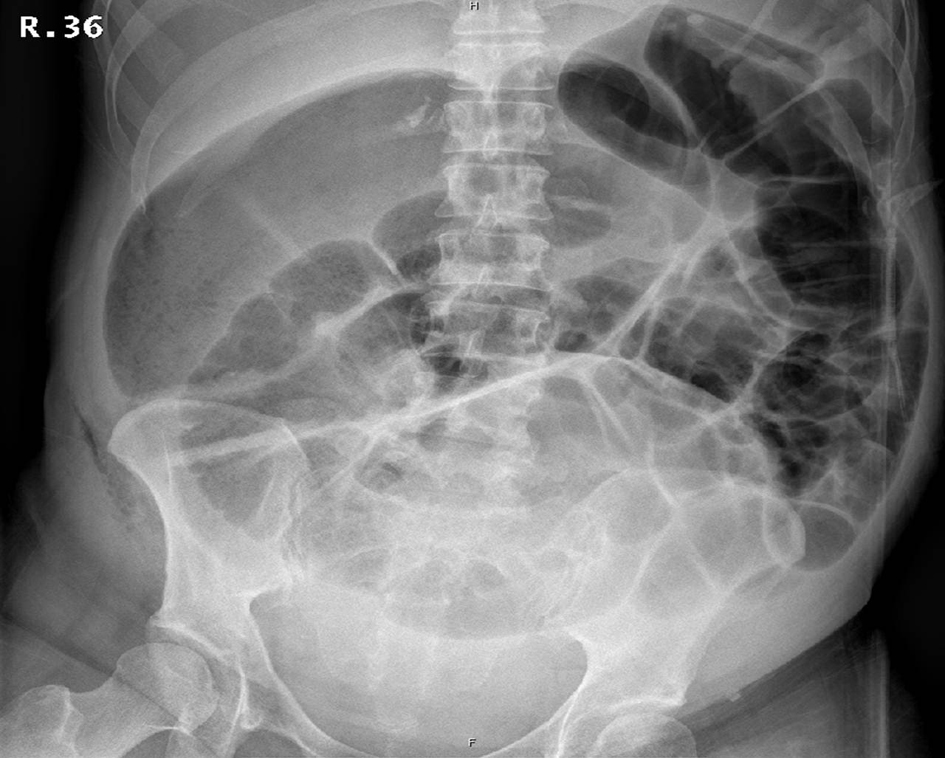
Figure 1. Abdominal X-ray shows grossly dilated bowel loops > 9 cm.
| Journal of Medical Cases, ISSN 1923-4155 print, 1923-4163 online, Open Access |
| Article copyright, the authors; Journal compilation copyright, J Med Cases and Elmer Press Inc |
| Journal website http://www.journalmc.org |
Case Report
Volume 4, Number 2, February 2013, pages 99-101
Surgical Management of Ogilvie’s Syndrome Post Caesarean Section
Figures