

| Journal of Medical Cases, ISSN 1923-4155 print, 1923-4163 online, Open Access |
| Article copyright, the authors; Journal compilation copyright, J Med Cases and Elmer Press Inc |
| Journal website http://www.journalmc.org |
Case Report
Volume 4, Number 5, May 2013, pages 284-287
Intra-Articular Dislocation of Patella Following Patellofemoral Arthroplasty
Colin S Holtona, b, Andrew P Cohena
aDepartment of Trauma & Orthopaedics, Pinderfields General Hospital, Wakefield, UK
bCorresponding author: Colin Holton, 319 Lidgett Lane, Leeds, West Yorkshire, LS17 6PD, UK
Manuscript accepted for publication November 16, 2012
Short title: Intra-Articular Dislocation of Patella
doi: https://doi.org/10.4021/jmc968e
| Abstract | ▴Top |
We present the first documented case of intra-articular dislocation of the patella following a patellofemoral arthroplasty on a 43-year-old male. This complication, caused by excessive flexion of the knee, was treated by biplanar manipulation under anaesthesia to effect a closed reduction and restoration of a safe range of motion.
Keywords: Dislocation; Patellofemoral arthroplasty; Biplanar manipulation
| Introduction | ▴Top |
Complications following arthroplasty of the patellofemoral joint are rare, but potentially catastrophic. These can include fracture of the patella or femur through over-resection, maltracking and component dissociation. Stiffness can result from component malposition, usually from inadequate resection of the femur or patella, resulting in ‘overstuffing’. Instability can result from inadequate soft tissue balancing or component malpositioning, commonly resulting in persistent coronal or axial plane malalignment and possible lateral subluxation or dislocation. Instability in the sagittal plane is much less common, and previously intra-articular patella dislocation has only been described in native knee joints caused by osteophyte formation. The patella can rotate in a horizontal or vertical axis and then becomes locked within the knee.
In this case, the patella dislocated inferiorly during excessive flexion resulting in a knee locked in flexion. The patella was reduced by manipulation under anaesthesia with no short-term complications.
| Case Report | ▴Top |
A 43-year-old male presented to Accident and Emergency with a painful knee locked in flexion. He had undergone a patellofemoral joint arthroplasty three months prior to this presentation. This procedure was performed at a different hospital in the region, the indications being intractable anterior knee pain with clinical and radiological features of isolated patellofemoral arthropathy. An FPV (Wright Cremascoli) patellofemoral arthroplasty had been implanted with size 1 trochlea and size 35 patella components. Pre-operatively, his range of motion had been well preserved at 0 to 130 degrees of flexion, and he consequently rapidly regained a satisfactory range of post-operative knee flexion, documented at 0 to 120 degrees by six weeks. No post-operative complications were noted.
Whilst cutting his toenails with his knee flexed to 130 degrees, he experienced severe anterior knee pain and was unable to straighten his right knee or weight-bear.
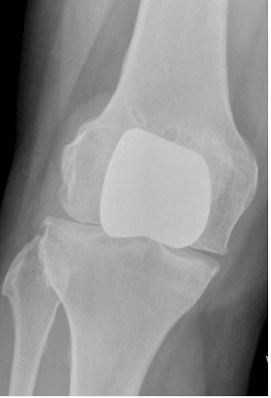
Examination revealed his right knee to be locked in 85 degrees of flexion, and radiographs confirmed a horizontal intra-articular dislocation of his patella (Fig 1, 2).
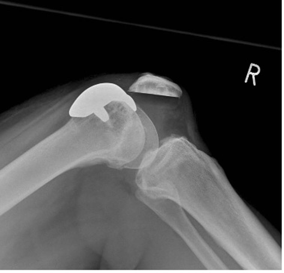 Click for large image | Figure 1. Lateral radiograph right knee, illustrating patella dislocation. |
 Click for large image | Figure 2. Meckel’s diverticulum after opening of the hernia sac. |
Attempted reduction in accident and emergency under entonox and morphine was unsuccessful.
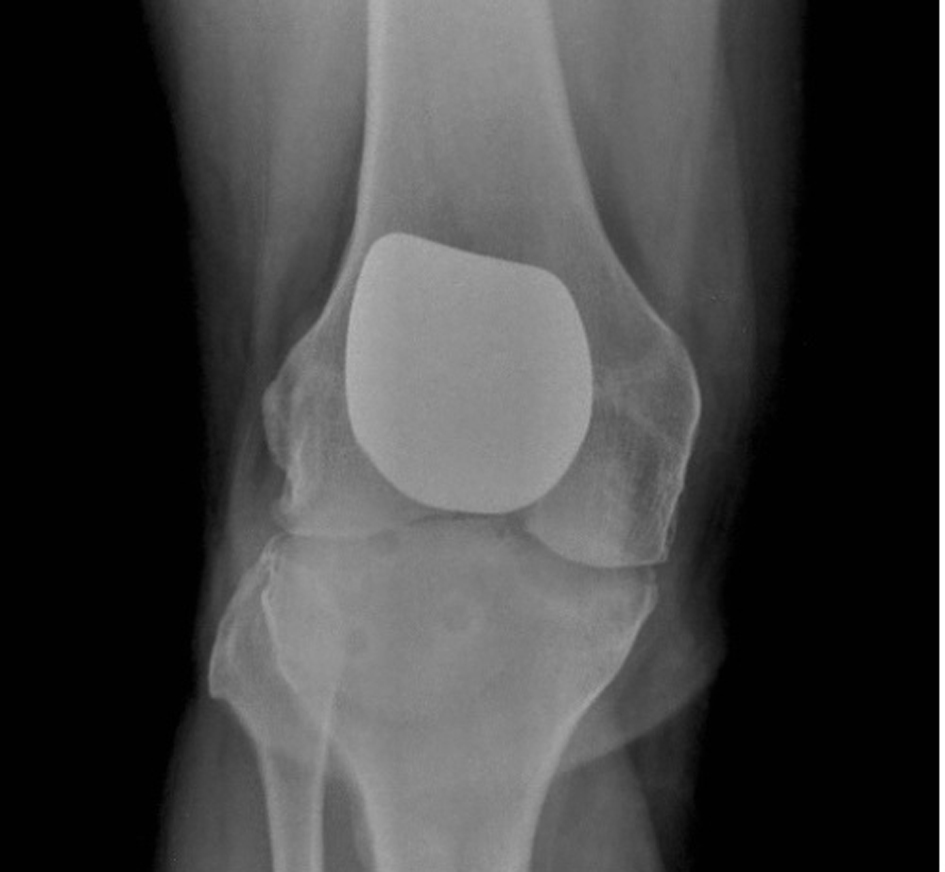
After admission and assessment on the trauma unit, consent was obtained for manipulation under anaesthesia and possible open reduction of dislocated patella. A general anaesthetic was administered, and closed reduction was achieved through a combination of hyper-flexion and internal rotation of his tibia to disengage his patella button from the femoral component (Fig. 3-5).
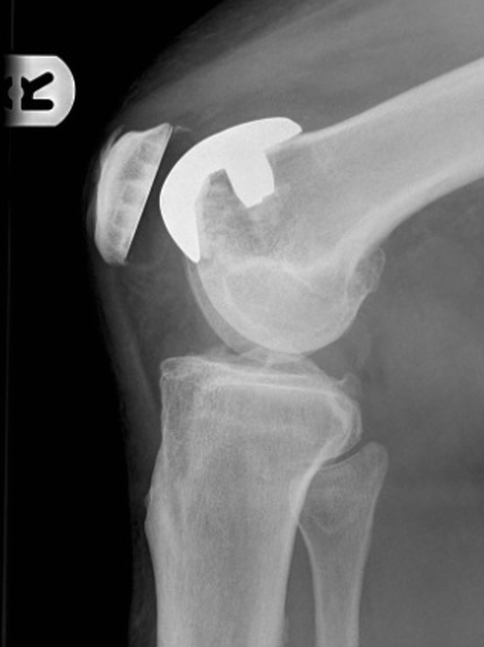 Click for large image | Figure 3. Lateral radiograph right knee, post reduction. |
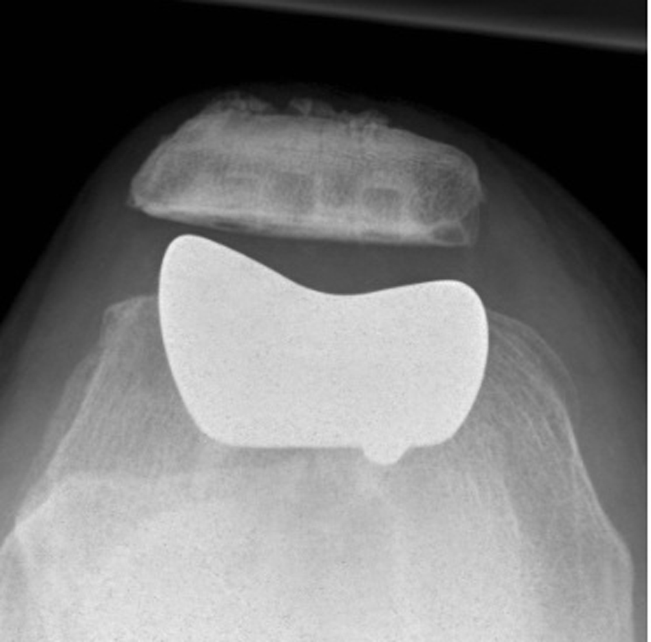 Click for large image | Figure 4. Skyline radiograph right knee, post reduction. |
 Click for large image | Figure 5. AP radiograph right knee, post reduction. |
Post-operatively he underwent splitting in a universal knee brace in full extension for 2 weeks followed by 4 weeks of controlled flexion of his knee, gradually increasing his range of flexion in 45 degree increments to 90 degrees at 6 weeks. Thereafter, his brace was discarded and increased flexion permitted, avoiding tibiofemoral rotation and activities involving flexion beyond 120 degrees.
At 3 years follow-up, his knee function has remained satisfactory. On examination he has 0 degrees to 120 degrees with no pain. No further episodes of dislocation and he has returned to normal activities of daily living.
| Discussion | ▴Top |
Long-term results following patellofemoral arthroplasty have reported complications including infection, fracture, component loosening, catching of prosthesis, mal-alignment and progression of disease requiring conversion to total knee arthroplasty (TKA) [1, 2]. Catching of the prosthesis noted in 2 cases highlights the need for correct placement of both components [2]. Although catching and mal-alignment have been noted as complications following patellofemoral arthroplasty, no reported cases of intra-articular dislocation following this procedure have been documented in the literature.
Commonly the patella dislocates to the lateral side with simple reduction usually possible without surgical intervention. Intra-articular dislocation of a native patella is very rare with only forty-seven reported cases in the literature. It dislocates in a horizontal or vertical axis of rotation. This condition was first reported by Midelfart in 1887 [3]. Only 8 vertical intra-articular dislocations have been reported in the literature [4-6] and occur less frequently than horizontal dislocations. Vertical intra-articular dislocation has most commonly been reported in adolescent boys [6]. It is thought to be due to ligament laxity at this age group which allows greater mobility of the patella which dislocates rather than fractures which are seen in the older population [7].
The commoner horizontal dislocations can be further subdivided into superior or inferior types. Superior horizontal intra-articular dislocations are rarer than the inferior type, and in these cases the mechanism is usually impact to the inferior patella pole [4]. This type of dislocation usually affects the elderly population with arthritic knees in which inferior patella osteophytes become interlocked with supra-condylar osteophytes [4, 8-10]. Inferior horizontal intra-articular dislocations normally occur due to the proximal pole of the patella being avulsed from the quadriceps tendon, lodging in the intra-condylar notch with the plane of the patella being parallel to the surface of the tibia [11, 12]. It is possible for the patella tendon to be detached and therefore allow the patella to be locked intra-articularly with the articular surface of the patella directed superiorly [12].
Native intra-articular dislocations have been treated by both open and closed methods. Most reported cases have required open reduction with only 12 reduced closed [7]. Murakami proposed that closed reduction was possible but depended on the degree of locking of the patella within the joint [13]. Choudhary comments that the method of reduction should be individualised to each patient depending on the degree of rotation of the patella. He further comments that if the patella is rotated beyond 90 degrees then open reduction should be performed [7].
This case is the first reported in a prosthetic patellofemoral joint. The likely cause of the dislocation was different to that of the native joint. In contrast to either impinging osteophtes or mechanical disruption, the mechanism of dislocation in this case appears to be catching of the proximal aspect of the resurfaced patella against the inferior aspect (tail) of the trochlea groove component. This may relate to inadequate recession of the trochlea groove component, or eccentric placement of the patella component leaving an ‘overhang’ of the patella component proximally. As in this case, inferior dislocation can then occur if sufficient flexion allows the proximal patella to migrate distally to the tail of the trochlea groove component, thus causing engagement of the two components with subsequent locking of the knee in flexion. In this case, reduction was achieved by biplanar manipulation, increasing flexion to disengage the components in the sagittal plane before correction of malrotation, thereby allowing the knee to be extended safely. Subsequent restrictions in rehabilitation allowed the knee to stabilise, without recurrence of instability. This may relate to the gradual formation of ‘meniscoid’ soft tissue around the patella component, commonly seen during revision arthroplasties, which may have minimised the chances of impingement of the patella and trochlea groove components. This case emphasises the need for correct placement of the components during patellofemoral arthroplasty. Younger patients with early disease frequently regain an enhanced range of flexion, and intra-operative analysis must ensure that catching of the components is avoided. Mal-position of either can lead to intra-articular dislocation, in this case possibly due to either an overhanging patella button or an inadequately recessed trochlea groove component. Without an open procedure, it is difficult to clarify the particular abnormality, though radiographic analysis suggests the patella component to be the correct size without significant proximal overhang. This suggests that the trochlea groove component may have been inadequately recessed or oversized allowing impingement.
In this case, a closed reduction was possible, with prevention of recurrent instability through a bracing programme and avoidance of excessive flexion.
Conflicts of Interest
None.
| References | ▴Top |
- Tauro B, Ackroyd CE, Newman JH, Shah NA. The Lubinus patellofemoral arthroplasty. A five- to ten-year prospective study. J Bone Joint Surg Br. 2001;83(5):696-701.
doi pubmed - Kooijman HJ, Driessen AP, van Horn JR. Long-term results of patellofemoral arthroplasty. A report of 56 arthroplasties with 17 years of follow-up. J Bone Joint Surg Br. 2003;85(6):836-840.
pubmed - Midelfart V. En sjelden luxation of patella. Norsk Mah Laeg 1887; 4: 588.
- Garner JP, Pike JM, George CD. Intra-articular dislocation of the patella: two cases and literature review. J Trauma. 1999;47(4):780-783.
doi pubmed - Alioto RJ, Kates S. Intra-articular vertical dislocation of the patella: a case report of an irreducible patellar dislocation and unique surgical technique. J Trauma. 1994;36(2):282-284.
doi - Nanda R, Yadav RS, Thakur M. Intra-articular dislocation of the patella. J Trauma. 2000;48(1):159-160.
doi pubmed - Choudhary RK, Tice JW. Intra-articular dislocation of the patella with incomplete rotation—two case reports and a review of the literature. Knee. 2004;11(2):125-127.
doi - Rao JP, Meese MA. Irreducible superior dislocation of the patella requiring open reduction. Am J Orthop (Belle Mead NJ). 1997;26(7):486-488.
pubmed - Friden T. A case of superior dislocation of the patella. Acta Orthop Scand. 1987;58(4):429-430.
doi pubmed - McWilliams TG, Binns MS. A locked knee in extension: a complication of a degenerate knee with patella alta. J Bone Joint Surg Br. 2000;82(6):890.
doi - Sarkar SD. Central dislocations of the patella. J Trauma. 1981;21(5):409-410.
doi pubmed - Nsouli AZ, Nahabedian AM. Intra-articular dislocation of the patella. J Trauma. 1988;28(2):256-258.
doi - Murakami Y. Intra-articular dislocation of the patella. A case report. Clin Orthop Relat Res. 1982;171:137-139.
This is an open-access article distributed under the terms of the Creative Commons Attribution License, which permits unrestricted use, distribution, and reproduction in any medium, provided the original work is properly cited.
Journal of Medical Cases is published by Elmer Press Inc.