

| Journal of Medical Cases, ISSN 1923-4155 print, 1923-4163 online, Open Access |
| Article copyright, the authors; Journal compilation copyright, J Med Cases and Elmer Press Inc |
| Journal website http://www.journalmc.org |
Case Report
Volume 5, Number 3, March 2014, pages 155-156
Unusual Two Foreign Bodies in Rectum: Case Report
Ugur Dogana, Umit Koca, Ismail Gomcelia, Mani Habibia, c, Tarik Gandi Cincinb, Nurullah Bulbullera
aGeneral Surgery Department, Antalya Training and Research Hospital, Antalya, Turkey
bGeneral Surgery Department, Iskenderun State Hospital, Iskenderun, Turkey
cCorresponding author: Mani Habibi, Department of General Surgery, Antalya Training and Research Hospital, Antalya, Turkey
Manuscript accepted for publication January 24, 2014
Short title: Foreign Bodies in Rectum
doi: https://doi.org/10.14740/jmc1672w
| Abstract | ▴Top |
In recent years, foreign bodies (FBs) in the rectum area are increasingly seen and complications that arise from this make up one of the most important issues in emergency surgery. Anorectal FB may result either from an orally ingested object that becomes impacted or more commonly due to insertion of objects through the anal canal. Unless there is a sign of peritonitis, the use of less invasive techniques is preferred for FB retrieval. Here, we present a case of a 54-year-old male patient with two FBs in rectum (stones), introduced as sexual perversion.
Keywords: Foreign bodies; Rectum; Complication
| Introduction | ▴Top |
In recent years, the incidence of patients presenting with rectal foreign bodies (FBs) has been increasing. Anorectal FB may result either from an orally ingested object that becomes impacted or more commonly due to insertion of objects through the anal canal. While mouth reception types are mostly encountered in children with mental disabilities, rectal insertive types are mostly used in middle-aged men for sexual stimulation [1, 2]. As a rectal FB is often associated with anal sexual behavior disorders, seeking treatment for its retrieval is often unpleasant and embarrassing for patients. FB may cause serious surgical issues due to its complications.
| Case Report | ▴Top |
We present here the case of a 54-year-old man admitted to the emergency department with rectal pain and constipation. The patient reported that 12 h prior to admission, he had used stones to clean his anus after defecation, and that these stones had become impacted in the anal canal. On digital rectal examination, FB was found to be palpated at 8 cm from the anal verge. There were two FBs seen on plain X-ray of abdomen around rectosigmoid region (Fig. 1). In lithotomy position forceps were used successfully to retrieve the FB after the anal dilatation under mask anesthesia. Stones measured 11 cm and 13 cm in length (Fig. 2). The patient was discharged after 24 h observation.
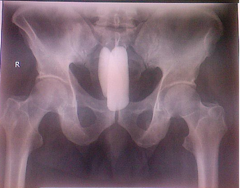 Click for large image | Figure 1. Two FBs seen on plain X-ray of abdomen. |
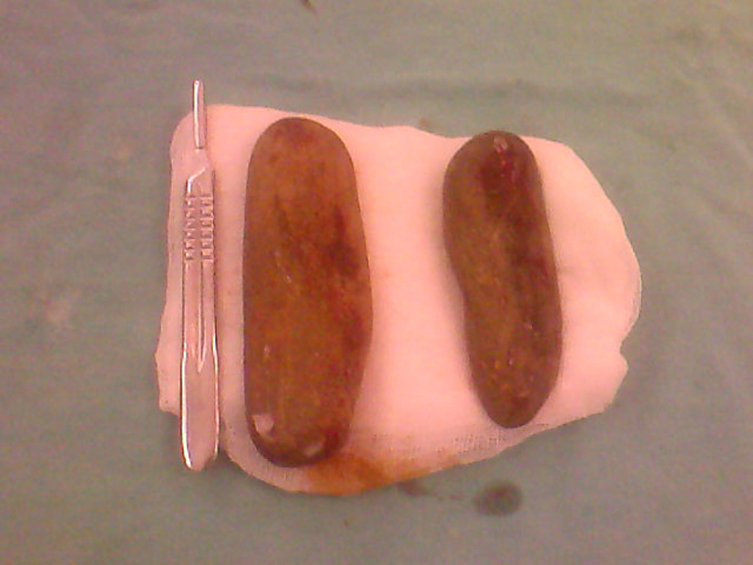 Click for large image | Figure 2. Two stones retrieved from the rectum. |
| Discussion | ▴Top |
Recently cases of rectal FBs have been encountered more frequently in clinical practice. Various objects have been described as retained rectal FBs, such as bottle, eggplant, vibrator used for sexual or erotic purposes, thermometer while applying medical treatment (as accident), irrigation catheter and enema container. Mouth reception types include dental prosthesis, needle and wood stick. Rectal pain and bleeding are the two main symptoms associated with rectal FB. It may cause perforation and acute abdominal pain [3]. Sometimes, it can also be related with rectal and vulvar abscess. Migration of FB to urinary and genital tract has been reported. Ooi et al reported that atypical gender behavior, lax anal sphincters and bloody or mucoid rectal discharge are the factors which raise the suspicion for FB [4].
The majority of FBs can be palpated at the middle rectum. If it is not possible to palpate, endoscopic and radiologic investigation should be performed. Plain abdominal X-rays are indicated in almost all cases; CT scans should be reserved for those with potential sepsis or equivocal peritoneal signs [5]. Unless there is a sign of peritonitis, it may be appropriate to wait several hours for spontaneous removal. In all cases, an appropriate tetanus prophylaxis and a prophylactic antibiotic are indicated. The genitourinary tract should also be examined for trauma and, if indicated, treatment. If possible, retrieval of FBs below the rectosigmoid region should be conducted transanally under sedation in the lithotomy position, using forceps and anal ecartors if necessary. If the patient cannot tolerate the pain, spinal or general anesthesia may be administered.
Often, use of the hand is the most convenient and easiest means of retrieving FBs from the rectum. In some cases, use of a Foley catheter, colonoscope, or Sengstaken-Blakemore tube may be necessary for retrieval. The main principle is to retrieve the FB transanally; if fails, it is better to retrieve it transanally after laparotomy with the assistance of the milking maneuver [6]. Bak et al described a novel approach to retrieval and removal of a rectal FB utilizing a single-incision laparoscopic surgery port [7]. Colotomy is the last option in the retrieval of FBs. If blood is detected during rectal examination, a sign of mucosal injury, the grade of injury must be evaluated after retrieval by performance of rectosigmoidoscopy or contrast studies.
It has been reported that primary repair, proximal loop colostomy, sigmoid end-colostomy and the Hartmann procedure, in combination with administration of wide-spectrum antibiotics according to the severity of peritoneal contamination, can be performed for the treatment of perforation. The mortality and morbidity rates of patients presenting with perforation above the peritoneal reflection have been reported to range from 2.5 to 20.0% and 20.0 to 40.0%, respectively [8, 9].
Conclusion
It is important to consider that FBs in the rectosigmoid region may cause perforation, peritonitis, pararectal abscess, fistulisation and genitourinary system injuries. It is also important to remember that complications may occur during FB retrieval. Thus, unless there is a sign of peritonitis, the use of less invasive techniques is preferred for FB retrieval.
| References | ▴Top |
- Fry RD. Anorectal trauma and foreign bodies. Surg Clin North Am. 1994;74(6):1491-1505.
pubmed - Atila K, Sokmen S, Astarcioglu H, Canda E. [Rectal foreign bodies: a report of four cases]. Ulus Travma Acil Cerrahi Derg. 2004;10(4):253-256.
pubmed - Goh BK, Chow PK, Quah HM, Ong HS, Eu KW, Ooi LL, Wong WK. Perforation of the gastrointestinal tract secondary to ingestion of foreign bodies. World J Surg. 2006;30(3):372-377.
doi pubmed - Ooi BS, Ho YH, Eu KW, Nyam D, Leong A, Seow-Choen F. Management of anorectal foreign bodies: a cause of obscure anal pain. Aust N Z J Surg. 1998;68(12):852-855.
doi pubmed - Desai B. Visual diagnosis: Rectal foreign body: A primer for emergency physicians. Int J Emerg Med. 2011;4:73.
doi pubmed - Singaporewalla RM, Tan DE, Tan TK. Use of endoscopic snare to extract a large rectosigmoid foreign body with review of literature. Surg Laparosc Endosc Percutan Tech. 2007;17(2):145-148.
doi pubmed - Bak Y, Merriam M, Neff M, Berg DA. Novel approach to rectal foreign body extraction. JSLS. 2013;17(2):342-345.
doi pubmed - Yildiz SY, Kendirci M, Akbulut S, Ciftci A, Turgut HT, Hengirmen S. Colorectal emergencies associated with penetrating or retained foreign bodies. World J Emerg Surg. 2013;8(1):25.
doi pubmed - Kasotakis G, Roediger L, Mittal S. Rectal foreign bodies: A case report and review of the literature. Int J Surg Case Rep. 2012;3(3):111-115.
doi pubmed
This is an open-access article distributed under the terms of the Creative Commons Attribution License, which permits unrestricted use, distribution, and reproduction in any medium, provided the original work is properly cited.
Journal of Medical Cases is published by Elmer Press Inc.