

| Journal of Medical Cases, ISSN 1923-4155 print, 1923-4163 online, Open Access |
| Article copyright, the authors; Journal compilation copyright, J Med Cases and Elmer Press Inc |
| Journal website https://www.journalmc.org |
Case Report
Volume 15, Number 1, January 2024, pages 15-19

An Uncommon Instance of Tubal Pregnancy With a Vital Fetus at the Thirteenth Week of Gestation
Vlora Ademi Ibishia, b, c ![]() , Naser Rafunab
, Naser Rafunab ![]() , Kaltrina Kolgecib
, Kaltrina Kolgecib ![]()
aMedical Faculty, University of Prishtina “Hasan Prishtina”, Prishtina 10000, Kosovo
bObstetrics and Gynecology Clinic, University Clinical Center of Kosova, Prishtina 10000, Kosovo
cCorresponding Author: Vlora Ademi Ibishi, Medical Faculty, University of Prishtina “Hasan Prishtina”, Prishtina 10000, Kosovo
Manuscript submitted November 22, 2023, accepted January 11, 2024, published online January 28, 2024
Short title: Tubal Pregnancy at the 13th Week of Gestation
doi: https://doi.org/10.14740/jmc4173
| Abstract | ▴Top |
Ectopic pregnancy, characterized by the aberrant implantation of blastocysts outside the uterine endometrial lining, typically occurs within the fallopian tube. Clinical presentation of tubal pregnancy ranges from asymptomatic cases to a spectrum of symptoms, including amenorrhea, pelvic pain, vaginal bleeding, and, critically, hemorrhagic shock resulting from tubal rupture. Utilizing serum beta-human chorionic gonadotropin levels and ultrasound examinations is pivotal in confirming the diagnosis. Diagnosing tubal pregnancy during the first trimester is feasible and crucial to prevent rupture, yet reports of fatal cases persist due to delays in diagnosis or misdiagnosis. This paper presents a unique case of an advanced tubal pregnancy at 13 weeks of gestation, featuring a viable fetus. This case underscores the importance of timely and accurate diagnosis to avoid life-threatening complications. It highlights the critical need for heightened medical vigilance and continuous education among healthcare professionals in managing ectopic pregnancies effectively.
Keywords: Ectopic pregnancy; Hemoperitoneum; Tubal rupture; Ultrasonography
| Introduction | ▴Top |
Ectopic pregnancy, characterized by the abnormal implantation of blastocysts outside the uterine endometrial lining, represents a potentially life-threatening condition with a prevalence of 1-2% in all pregnancies [1]. Predominantly, 96% of these cases manifest as tubal pregnancies, where the ectopic pregnancy develops within the fallopian tubes [2]. Comparatively, the ovaries, abdominal cavity, and cervix uteri are exceedingly rare sites for pathologic implantation [3]. Tubal pregnancies typically become symptomatic during the first trimester of pregnancy due to the stretching and erosion of the fallopian tube walls as a result of blastocyst development. Clinically, the condition is characterized by the classic triad of manifestations - amenorrhea, low abdominal pain, and vaginal bleeding.
Moreover, diagnosis is refined through evaluating serum beta-human chorionic gonadotropin (hCG) levels and using ultrasound examinations. Timely detection and management, ideally within the sixth to ninth week of gestation, are pivotal for preventing tubal rupture [4]. The longer the condition remains undiagnosed and untreated, the greater the risk of tubal rupture, leading to internal bleeding and shock [5]. The timing of rupture varies among individual cases. However, it generally occurs around 7.2 gestational weeks, with a variance of ± 2.2 weeks, representing a severe hazard to the patient’s health and life [6].
The case under consideration pertains to an exceedingly rare occurrence of a tubal pregnancy that remained untreated and unruptured until the 13th week of gestation, housing a vital fetus and thereby placing the patient in a state of extreme risk.
| Case Report | ▴Top |
Investigations
A 36-year-old patient, gravida 5, para 4, presented herself at the Obstetrics and Gynecology Clinic, complaining of lower abdominal pain and vaginal bleeding. At the time, she was in her 13th week of pregnancy, and her last gynecological examination, conducted 2 months earlier, had confirmed a 5-week intrauterine pregnancy. In response to inquiries about her irregular check-ups, the patient expressed her belief that her pregnancy was progressing normally, and she considered additional appointments unnecessary. Upon physical examination, the patient displayed stable vital signs, including a blood pressure of 100/70 mm Hg and a pulse rate of 80 beats per minute. Palpation revealed a firm and sensitive lower abdomen. Gynecological examination revealed a uterus smaller than anticipated for the given gestational age, a sensitive uterus with pain originating from the left adnexa, a closed cervical ostium sensitive to manipulation, and slight uterine bleeding. Initial laboratory results indicated a hemoglobin level of 10.5 g/dL (normal range 12 - 16 g/dL) and a hematocrit level of 31% (normal range 36-48%).
Diagnosis
Subsequent pelvic ultrasound revealed an empty uterine cavity with an endometrial thickness of 9 mm. Remarkably, to the left and outside of the uterus, a live fetus with biometric data reflecting a crown-rump length (CRL) of 64 mm, biparietal diameter (BPD) of 21 mm, and femur length (FL) of 10 mm, corresponding to the 13th week of gestation, was observed (Fig. 1a-c). The fetal cardiac activity was recorded, as shown in Figure 1a, with a yellow arrow.
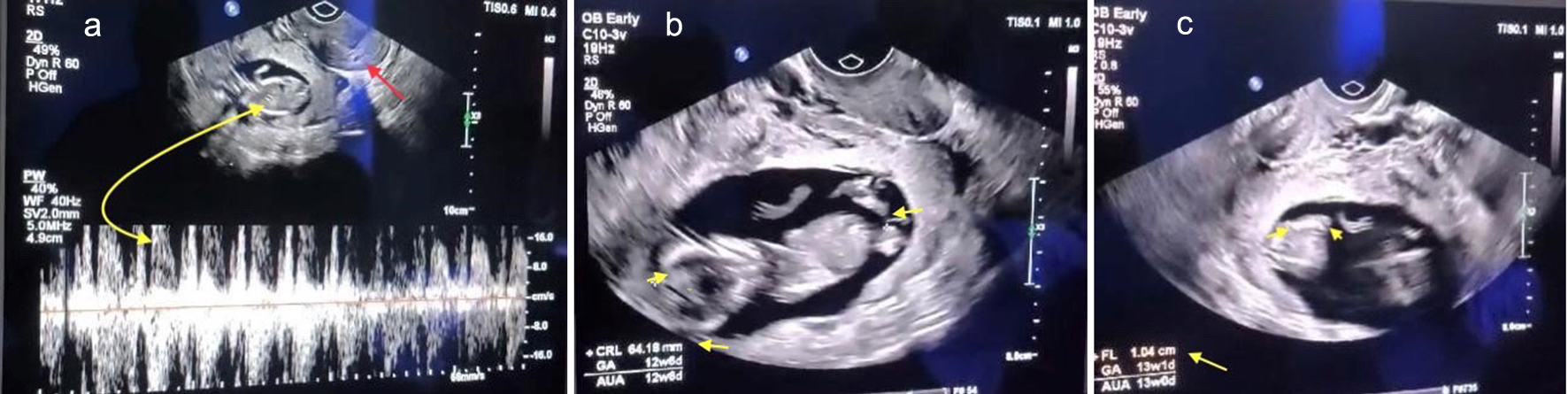 Click for large image | Figure 1. (a) Transvaginal sonographic view of tubal pregnancy depicting an empty uterus (red arrow) with a viable ectopic pregnancy (yellow arrow). (b) Yellow arrowhead points reflecting the crown rump length of the fetus. (c) Yellow arrow points representing the femur length corresponding to a fetus at the 13th week of gestation. |
Nonetheless, the presence of a substantial volume of amniotic fluid and the observation of free fetal movements led to a sonographic interpretation suggestive of an intact abdominal ectopic pregnancy. A minimal amount of free fluid was noted in Douglas’s pouch, while Morrison’s pouch was free of fluid. While preparing the patient for a magnetic resonance imaging (MRI) scan to ascertain the precise localization of the fetus, a rapid deterioration in her overall health became evident, characterized by progressive hypotension. In response to this critical development, an immediate decision was made to proceed with an exploratory laparotomy. This course of action necessitated the ordering of three units of whole blood for intraoperative resuscitation, with the patient granting written consent before being expedited into the operating room.
Treatment
A substantial volume of blood within the peritoneal cavity was observed during the surgical intervention, as shown in Figure 2.
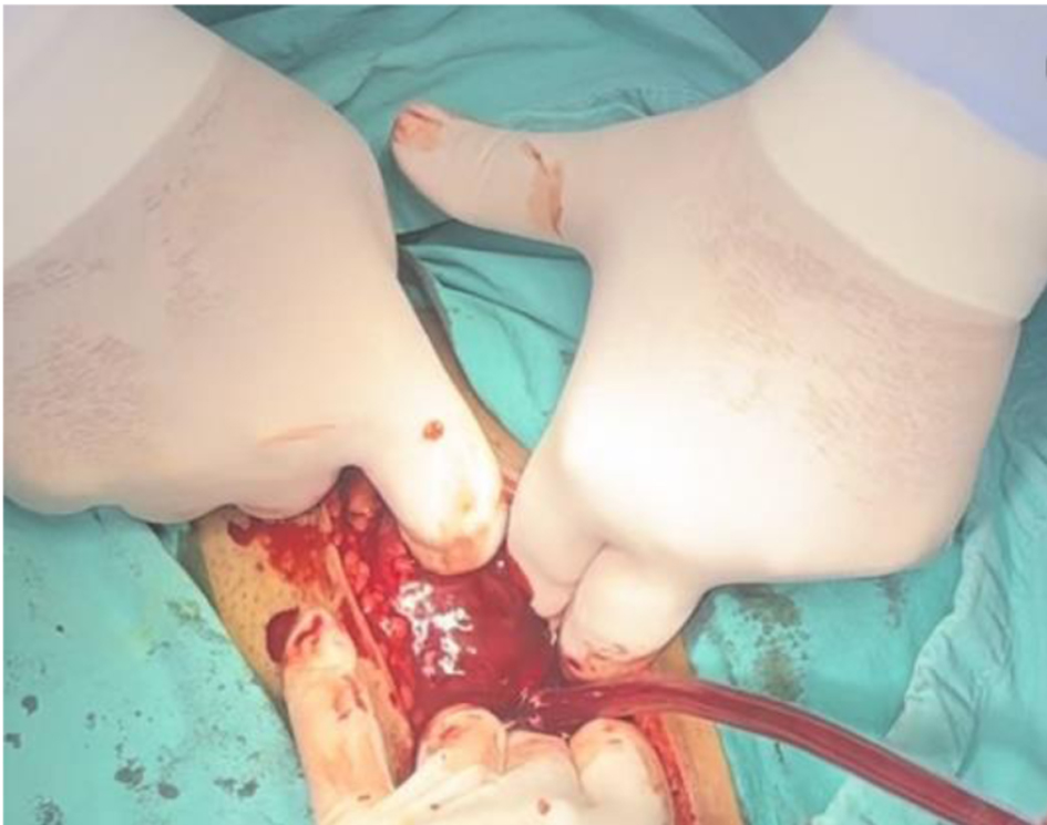 Click for large image | Figure 2. Intraoperative finding of hemoperitoneum resulting from hemorrhage in the context of an ectopic tubal pregnancy. |
After removing 900 mL of blood, the pelvic organs became distinctly discernible. Notably, the uterus, ovaries, and the right fallopian tube exhibited a normal appearance. Conversely, the left fallopian tube displayed a considerable enlargement similar in size to that of the uterus. It was associated with a moderate hemorrhage, as visually presented in Figure 3.
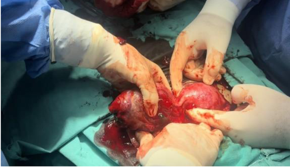 Click for large image | Figure 3. Enlarged left fallopian tube resulting from ectopic pregnancy, with the uterus displaying typical morphology. |
Promptly, it became evident that the 13-week ectopic pregnancy was confined within the left fallopian tube, necessitating a salpingectomy procedure, during which the patient received intraoperative transfusion support. Upon resection of the affected fallopian tube, an exploratory longitudinal incision was executed, revealing the presence of a 13-week-old fetus, placenta, and intact chorioamniotic membranes (Fig. 4).
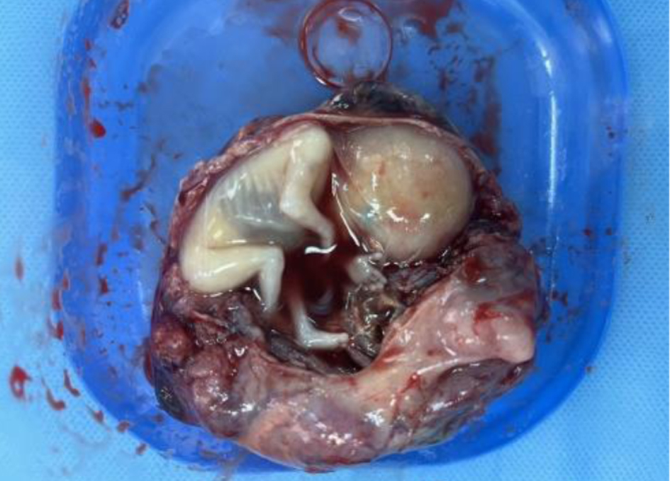 Click for large image | Figure 4. Macroscopic examination of a 13-week fetus, placenta, and chorioamniotic membranes within the resected fallopian tube. |
Follow-up and outcomes
Postoperatively, the patient’s recovery progressed without noteworthy incidents, leading to her discharge on the fourth day following the surgical procedure.
| Discussion | ▴Top |
Ectopic pregnancy is an important cause of obstetric morbidity and mortality in the first trimester, while cases of tubal pregnancy with a live fetus progressing into the second trimester, such as the one presented in this case, are rare and typically due to misdiagnosis or poor antenatal care.
An ectopic pregnancy is defined as any gestation implanting outside the uterine cavity, occurring in approximately 2% of all pregnancies. This condition adversely affects women’s reproductive health by decreasing fertility and increasing the risk of subsequent ectopic pregnancies. It is responsible for around 5-10% of deaths related to pregnancy [7, 8]. The vast majority of cases, approximately 96%, of ectopic pregnancies are primarily located in the uterine tubes, commonly referred to as tubal pregnancies. Other rare locations include the ovaries and abdominal cavity, often resulting from the extrusion of the ovum from the uterine tube. Lastly, constituting only 1% of ectopic pregnancies are intramural pregnancies and cesarean scar pregnancies [9]. Regardless of the location, ectopic pregnancy represents a life-threatening condition.
Various risk factors contribute to ectopic pregnancies, including pelvic inflammatory disease related to infections such as Chlamydia trachomatis, Neisseria gonorrhoeae, and Mycoplasma genitalium, previous ectopic pregnancies, tubal surgeries, advanced maternal age, smoking, multiparity, prior abortions, and assisted reproductive technologies [10, 11]. In our case, the associated risk factors were multiparity and advanced maternal age.
Clinical manifestations of tubal pregnancy span a spectrum, from asymptomatic presentations to a range of symptoms, including amenorrhea, pelvic pain, and vaginal bleeding. Untreated, it may culminate in the rupture of the fallopian tube, instigating internal bleeding and hemorrhagic shock. Tubal pregnancy has always been considered a complication of the first trimester of pregnancy, with a life-threatening rupture that often occurs around the seventh week of pregnancy [6].
Timely diagnosis and intervention in the early stages of the first trimester are crucial to prevent fallopian tube rupture and its complications. Diagnosis involves a combination of clinical symptoms, beta-hCG levels, and ultrasound imaging. Clinical signs of unruptured tubal pregnancies include lower abdominal pain and vaginal bleeding. These nonspecific symptoms may mimic appendicitis, ovarian cysts, or urolithiasis [12]. To avoid diagnostic delays, women of reproductive age with such symptoms should be considered as potential cases of ectopic pregnancy. They should undergo a pelvic ultrasound and beta-hCG levels assessment.
HCG level is a vital marker for diagnosing and monitoring early pregnancy. HCG levels above 1,500 - 2,000 mIU/mL, along with the absence of intrauterine pregnancy on ultrasound, strongly indicate ectopic pregnancy [13]. Assessing the increase of hCG level every 48 h provides insight into the development of the pregnancy. Pregnancies with an hCG level increase of less than 35% are likely to be ectopic pregnancies [14].
Ultrasound, particularly transvaginal sonography, is the imaging diagnostic modality of choice for ectopic pregnancies due to its availability, efficiency, accuracy, and non-invasive nature. Transvaginal sonography has shown high sensitivity (92.3%), specificity (75%), positive predictive value (96%), and accuracy (90%) in diagnosing suspected ectopic pregnancies, surpassing transabdominal sonography [15]. In developed countries, the availability of high-resolution ultrasound has enabled the detection of more than 80% of ectopic pregnancies before rupture, and more than 50% are diagnosed in asymptomatic women [16]. Diagnosis of ectopic pregnancy before rupture is of high importance because it enables either pharmacological or laparoscopic surgery, which is considered to be the gold standard and the most cost-effective surgical approach [17, 18].
In developing countries, the situation is quite different. Substandard antenatal care, low availability of high-resolution devices, and non-continuous training lower the possibility of a pre-rupture diagnosis.
Above the quality of the available tools, the determining factor in the diagnosing of an ectopic pregnancy is the clinician’s ability to recognize the sonographic features. Low abdominal pain and positive beta-hCG can be good indicators of ectopic pregnancy and are easy for a physician to recognize; however, to confirm it, a pelvic ultrasound is essential. Physicians must start by confirming the absence of an intrauterine pregnancy and then continue the sonographic examination to identify the characteristic features of an ectopic pregnancy. Notably significant are the formation of a pseudo-gestational sac and the presence of the “ring of fire”, both manifesting before rupture.
Approximately 20% of tubal pregnancies exhibit the formation of a pseudo-gestational sac, typically visible around the fifth week of pregnancy [19]. This pseudo-gestational sac comprises a small fluid collection within the uterus and, without adequate familiarity with its characteristics, may be misconstrued as a genuine gestational sac, potentially leading to misdiagnosis. Such a scenario was encountered in our case where, in a checkup in the fifth week of gestation, the pseudo-gestational sack was misinterpreted as being an intrauterine pregnancy. To avoid this misinterpretation, the visualization of an empty sac within the uterus cannot confirm an intrauterine pregnancy. Instead, confirmation should involve the visualization of embryonic landmarks like the yolk sac or fetal pole within the sac. In cases where these landmarks are absent, a follow-up appointment within a week is crucial to distinguish between a true gestational sac and a pseudo-gestational sac [20].
In approximately 71-90% of tubal ectopic pregnancies, a hyperechoic ring, commonly referred to as the “ring of fire”, encircles the central anechoic sac. Within this sac, structures such as the yolk sac, embryo, and even cardiac activity can frequently be discerned [21].
In cases in which tubal rupture occurs, typically around the 7.2 ± 2 weeks of gestation, diagnosis can be achieved by recognizing the clinical manifestation of hemorrhagic shock, positive beta-hCG, and ultrasonographic imaging of the peritoneal cavity displaying free fluid containing debris [22]. If these signs are recognized, a patient must undergo surgical intervention immediately to halt the internal bleeding and save the patient’s life.
Cases in which tubal pregnancies progress into the second trimester without rupture are very uncommon in literature. The case we are presenting is of a 13-week tubal pregnancy, which furthermore had a viable fetus, making it exceptionally rare. Archival research showed that a similar case in the past decade has yet to be presented in our tertiary institution, which handles over 10,000 births annually.
Ectopic pregnancies remain one of the leading causes of maternal death related to pregnancy, representing 5-10% of cases. Our case and others reported by authors all around the world show that ectopic pregnancies, even to this day, are allowed to progress undiagnosed or, worse, misdiagnosed, putting patients’ lives at risk [23-25].
Conclusion
However uncommon, tubal pregnancies progressing into the second trimester continue to be reported to this day, showing that delay or misdiagnosis of ectopic pregnancies is a persistent issue. Ultrasound examination is the ideal tool for recognizing ectopic pregnancies.
However, a physician should be familiar with its features, and continuous checkups must be performed for an early diagnosis. This allows for pharmacological treatment or laparoscopic surgery and decreases the chances of a tubal rupture. In cases where diagnosis is delayed until the second trimester, the patient’s life is at serious risk, and surgical intervention is the only method available and should be performed immediately.
Learning points
Second-trimester tubal pregnancy is a rare but life-threatening condition. Patients usually present with low abdominal pain and vaginal bleeding. Sonographic imaging of the fetus can often be deceiving and give the impression of an abdominal pregnancy. Once diagnosed, surgical intervention is necessary and must be undertaken immediately.
Persistent reports of second-trimester tubal pregnancy emphasize the need for improved antenatal care, medical vigilance, knowledge dissemination, and ongoing medical training to enhance healthcare professionals’ diagnostic abilities in managing ectopic pregnancies effectively.
Acknowledgments
None to declare.
Financial Disclosure
None to declare.
Conflict of Interest
None to declare.
Informed Consent
Written consent was obtained from the patient for the publication of this case and any accompanying images.
Author Contributions
Vlora Ademi Ibishi: conception and design. Vlora Ademi Ibishi and Naser Rafuna: manuscript writing. Vlora Ademi Ibishi and Kaltrina Kolgeci: data collection. Vlora Ademi Ibishi, Naser Rafuna and Kaltrina Kolgeci: review, corrections and approval of the manuscript.
Data Availability
The authors declare that data supporting the findings of this study are available within the article.
| References | ▴Top |
- Alkatout I, Honemeyer U, Strauss A, Tinelli A, Malvasi A, Jonat W, Mettler L, et al. Clinical diagnosis and treatment of ectopic pregnancy. Obstet Gynecol Surv. 2013;68(8):571-581.
doi pubmed - Panelli DM, Phillips CH, Brady PC. Incidence, diagnosis and management of tubal and nontubal ectopic pregnancies: a review. Fertil Res Pract. 2015;1:15.
doi pubmed pmc - Barnhart KT. Clinical practice. Ectopic pregnancy. N Engl J Med. 2009;361(4):379-387.
doi pubmed - Kumar V, Gupta J. Tubal ectopic pregnancy. BMJ Clin Evid. 2015;2015:1406.
pubmed pmc - Gari R, Abdulgader R, Abdulqader O. A live 13 weeks ruptured ectopic pregnancy: a case report. Cureus. 2020;12(10):e10993.
doi pubmed pmc - Fan YY, Liu YN, Mao XT, Fu Y. The prevalence of ectopic gestation: a five-year study of 1273 cases. Int J Gen Med. 2021;14:9657-9661.
doi pubmed pmc - Li C, Zhao WH, Zhu Q, Cao SJ, Ping H, Xi X, Qin GJ, et al. Risk factors for ectopic pregnancy: a multi-center case-control study. BMC Pregnancy Childbirth. 2015;15:187.
doi pubmed pmc - Houser M, Kandalaft N, Khati NJ. Ectopic pregnancy: a resident’s guide to imaging findings and diagnostic pitfalls. Emerg Radiol. 2022;29(1):161-172.
doi pubmed - Rana P, Kazmi I, Singh R, Afzal M, Al-Abbasi FA, Aseeri A, Singh R, et al. Ectopic pregnancy: a review. Arch Gynecol Obstet. 2013;288(4):747-757.
doi pubmed - Yuk JS, Kim YJ, Hur JY, Shin JH. Association between socioeconomic status and ectopic pregnancy rate in the Republic of Korea. Int J Gynaecol Obstet. 2013;122(2):104-107.
doi pubmed - Bouyer J, Coste J, Shojaei T, Pouly JL, Fernandez H, Gerbaud L, Job-Spira N. Risk factors for ectopic pregnancy: a comprehensive analysis based on a large case-control, population-based study in France. Am J Epidemiol. 2003;157(3):185-194.
doi pubmed - Hendriks E, Rosenberg R, Prine L. Ectopic pregnancy: diagnosis and management. Am Fam Physician. 2020;101(10):599-606.
pubmed - Nwabuobi C, Arlier S, Schatz F, Guzeloglu-Kayisli O, Lockwood CJ, Kayisli UA. hCG: biological functions and clinical applications. Int J Mol Sci. 2017;18(10):2037.
doi pubmed pmc - Seeber BE, Sammel MD, Guo W, Zhou L, Hummel A, Barnhart KT. Application of redefined human chorionic gonadotropin curves for the diagnosis of women at risk for ectopic pregnancy. Fertil Steril. 2006;86(2):454-459.
doi pubmed - Nahar MN, Quddus MA, Sattar A, Shirin M, Khatun A, Ahmed R, Sultana F. Comparison of transvaginal and transabdominal ultrasonography in the diagnosis of ectopic pregnancy. Bangladesh Med Res Counc Bull. 2013;39(3):104-108.
doi pubmed - Winder S, Reid S, Condous G. Ultrasound diagnosis of ectopic pregnancy. Australas J Ultrasound Med. 2011;14(2):29-33.
doi pubmed pmc - Orazulike NC, Konje JC. Diagnosis and management of ectopic pregnancy. Womens Health (Lond). 2013;9(4):373-385.
doi pubmed - Di Lorenzo G, Romano F, Mirenda G, Cracco F, Buonomo F, Stabile G, Facchin S, et al. “Nerve-sparing” laparoscopic treatment of parametrial ectopic pregnancy. Fertil Steril. 2021;116(4):1197-1199.
doi pubmed - Chanana C, Gupta N, Bansal I, Hooda K, Sharma P, Gupta M, Gandhi D, et al. Different sonographic faces of ectopic pregnancy. J Clin Imaging Sci. 2017;7:6.
doi pubmed pmc - Barnhart K, van Mello NM, Bourne T, Kirk E, Van Calster B, Bottomley C, Chung K, et al. Pregnancy of unknown location: a consensus statement of nomenclature, definitions, and outcome. Fertil Steril. 2011;95(3):857-866.
doi pubmed pmc - Rempen A. Vaginal sonography in ectopic pregnancy. A prospective evaluation. J Ultrasound Med. 1988;7(7):381-387.
doi pubmed - Lawani OL, Anozie OB, Ezeonu PO. Ectopic pregnancy: a life-threatening gynecological emergency. Int J Womens Health. 2013;5:515-521.
doi pubmed pmc - Khalil A, Saber A, Aljohani K, Khan M. The efficacy and success rate of methotrexate in the management of ectopic pregnancy. Cureus. 2022;14(7):e26737.
doi pubmed pmc - Ngene NC, Lunda O. Ectopic pregnancy in the ampulla of the fallopian tube at 16 gestational weeks: lessons from a case report. Afr Health Sci. 2020;20(4):1895-1897.
doi pubmed pmc - Ilea C, Ilie OD, Marcu OA, Stoian I, Doroftei B. The very first Romanian unruptured 13-weeks gestation tubal ectopic pregnancy. Medicina (Kaunas). 2022;58(9):1160.
doi pubmed pmc
This article is distributed under the terms of the Creative Commons Attribution Non-Commercial 4.0 International License, which permits unrestricted non-commercial use, distribution, and reproduction in any medium, provided the original work is properly cited.
Journal of Medical Cases is published by Elmer Press Inc.