

| Journal of Medical Cases, ISSN 1923-4155 print, 1923-4163 online, Open Access |
| Article copyright, the authors; Journal compilation copyright, J Med Cases and Elmer Press Inc |
| Journal website http://www.journalmc.org |
Case Report
Volume 3, Number 3, June 2012, pages 211-213
Expanding Growth and Pharyngeal Mass Formation in Elderly Patient With Esophageal Cancer: Unusual Manifestation
Takatsugu Yamamotoa, b, Koichiro Abea, Hajime Anjikia, Taro Ishiia, Yasushi Kuyamaa
aDepartment of Internal Medicine, Teikyo University School of Medicine, Tokyo, Japan
bCorresponding author: Takatsugu Yamamoto, Department of Internal Medicine, Teikyo University School of Medicine, 2-11-1 Kaga, Itabashi-ku, 173-8605, Tokyo, Japan
Manuscript accepted for publication February 23, 2012
Short title: Esophageal Cancer and Pharyngeal Mass
doi: https://doi.org/10.4021/jmc587w
| Abstract | ▴Top |
Esophageal squamous cell carcinoma has invasive nature itself, and infiltration to the surrounding organs is common. We experienced a case of esophageal cancer with unusual manifestation. Eighty-one-year-old female with 3 years history of cognitive disorder referred to our hospital because of dysphagia lasting for a month. The upper gastrointestinal endoscopy with thin scope revealed a white-colored, soft mass lesion occupying almost whole of the lower pharynx. The lesion connected to the thickened esophageal wall, and the scope could not get through due to stenosis at the upper esophagus. The computed tomography showed marked thickness of the esophageal wall at the upper to middle portion without direct invasion or fistula formation to the surrounding organs. The lesion had protruding parts at the both edges, shown as low-density area on computed tomography, indicating inflammatory change. Pathological examination of the biopsied specimen showed squamous cell carcinoma of the esophagus and necrotic tissue of the pharynx. The patient initiated radiation therapy after tracheotomy was made, but died due to pneumonia two months later.
Keywords: Esophageal cancer; Expanding growth; Pharyngeal mass
| Introduction | ▴Top |
Esophageal squamous cell carcinoma has invasive nature itself, and infiltration to the surrounding organs such as bronchus is common. We report on a rare case of esophageal cancer with unusual manifestation.
| Case Report | ▴Top |
An 81-year-old female with 3 years history of cognitive disorder referred to our hospital because of dysphagia lasting for a month. The upper gastrointestinal endoscopy with thin scope revealed a white-colored, movable, soft mass lesion occupying almost whole of the lower pharynx (Fig. 1). The lesion connected to the thickened esophageal wall, and the scope could not get through due to stenosis at the upper esophagus (Fig. 2). The computed tomography showed marked thickness of the esophageal wall at the upper to middle portion. The lesion had protruding parts at the both edges, shown as low-density area on computed tomography, indicating inflammatory change (Fig. 3-5). Pathological examination of the biopsied specimen showed squamous cell carcinoma of the esophagus and necrotic tissue of the pharynx (Fig. 6). The patient initiated radiation therapy after tracheotomy was made, but died due to pneumonia two months later.
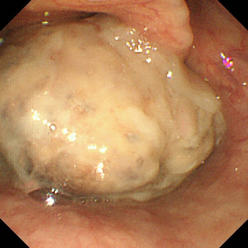 Click for large image | Figure 1. The upper gastrointestinal endoscopy with thin scope showed a white-colored, movable mass lesion occupying the lower pharynx. |
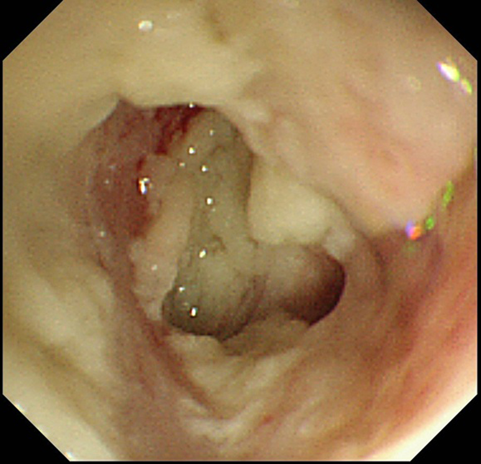 Click for large image | Figure 2. Stenosis was seen at the upper esophagus. The endoscope could not get through the stenosis. |
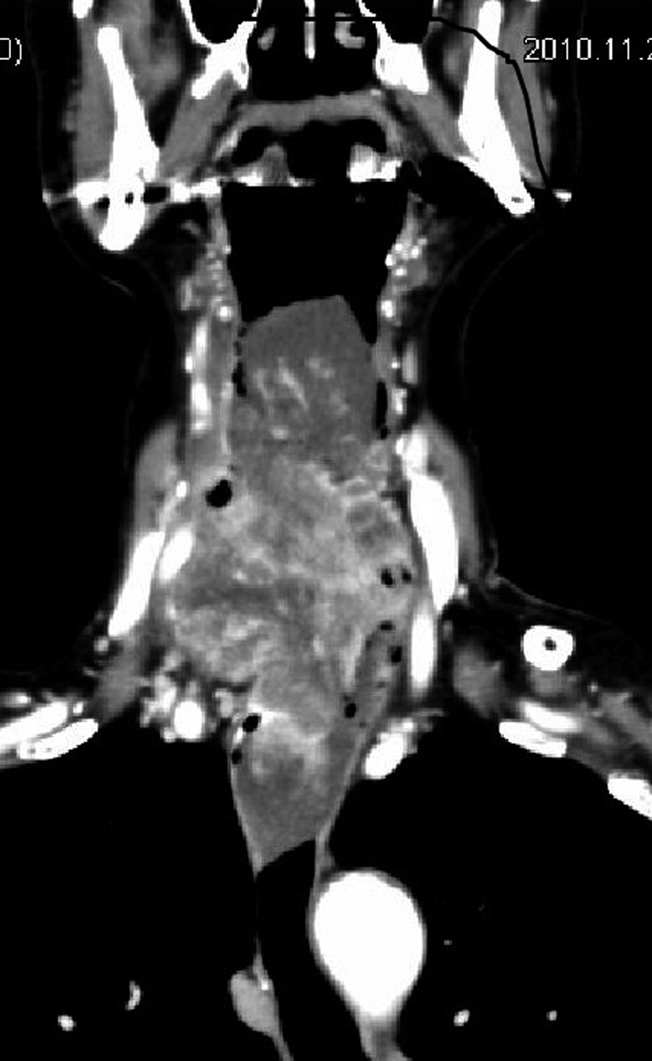 Click for large image | Figure 3. Computed tomography (frontal plane). A large mass occupied the lower pharynx. |
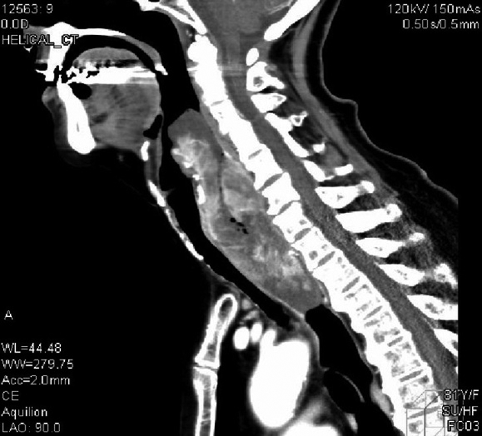 Click for large image | Figure 4. Computed tomography (sagittal plane). Marked wall thickness was seen at the upper to middle esophagus. The lesion had protruding parts at the both edges, shown as low-density area, indicating inflammatory change. |
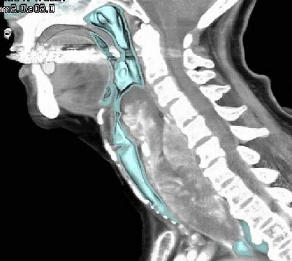 Click for large image | Figure 5. Computed tomography (sagittal plane with specific coloring). The border of the bronchus (showed as green area) looks clear and smooth. |
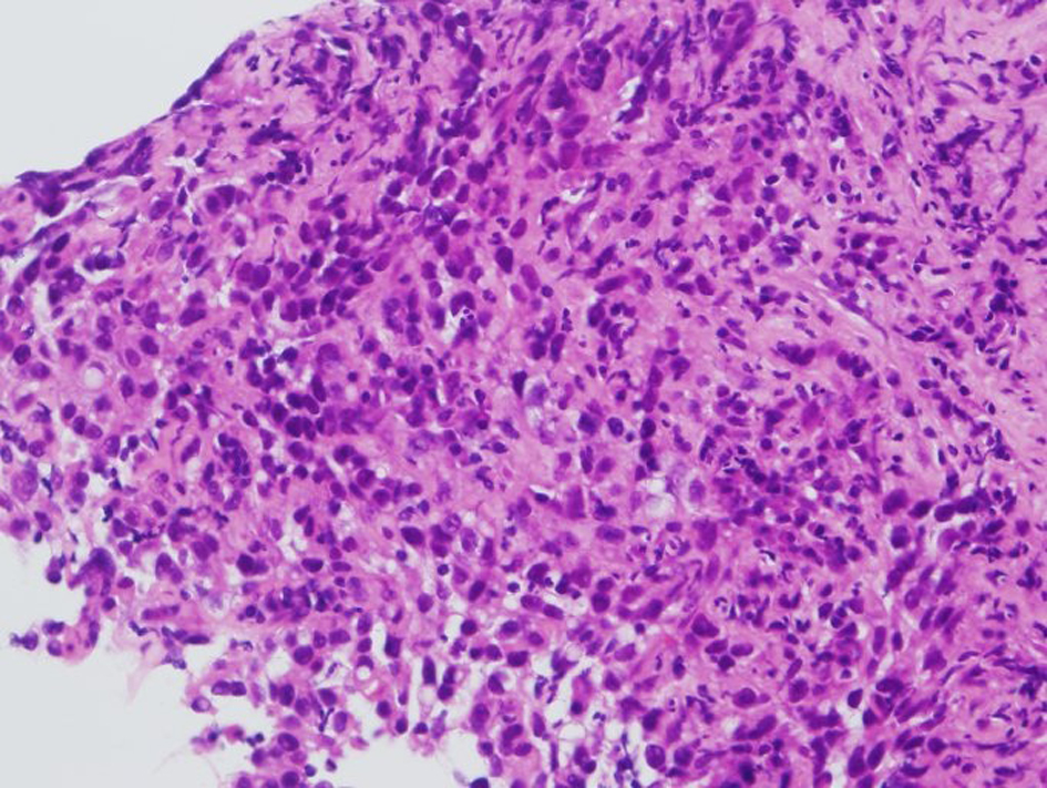 Click for large image | Figure 6. Pathological examination of the biopsied specimen showed squamous cell carcinoma of the esophagus (HE stain). |
| Discussion | ▴Top |
More than 90% of esophageal malignancies are squamous cell carcinoma in Japan [1]. Esophageal squamous cell carcinoma is known its invasive nature, and invasion to the surrounding tissues including respiratory organs and mediastinum is common clinically [2]. The present case was rare at this point of view. As shown in Figure 3-5, the margin of the tumor to the other organs was clear, suggesting that the tumor grew not invasively but expansively. Additionally, it was also rare that esophageal cancer formed pharyngeal mass protruding from esophageal wall. Clinicians should know the variation of growing pattern of esophageal cancer.
| References | ▴Top |
- Takubo K, Aida J, Sawabe M, Kurosumi M, Arima M, Fujishiro M, Arai T. Early squamous cell carcinoma of the oesophagus: the Japanese viewpoint. Histopathology. 2007;51(6):733-742.
pubmed doi - Wolf MC, Stahl M, Krause BJ, Bonavina L, Bruns C, Belka C, Zehentmayr F. Curative treatment of oesophageal carcinoma: current options and future developments. Radiat Oncol. 2011;6:55.
This is an open-access article distributed under the terms of the Creative Commons Attribution License, which permits unrestricted use, distribution, and reproduction in any medium, provided the original work is properly cited.
Journal of Medical Cases is published by Elmer Press Inc.